. 2019 Jul 19;10(1):3200.
doi: 10.1038/s41467-019-11162-4.

circTP63 functions as a ceRNA to promote lung squamous cell carcinoma progression by upregulating FOXM1

Affiliations

circTP63 functions as a ceRNA to promote lung squamous cell carcinoma progression by upregulating FOXM1

Zhuoan Cheng et al. Nat Commun. .

Abstract

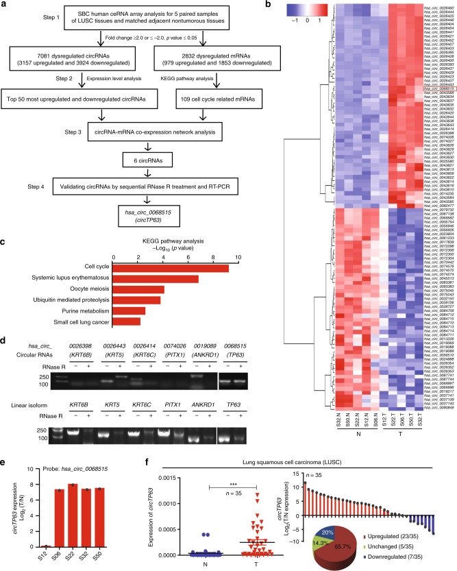
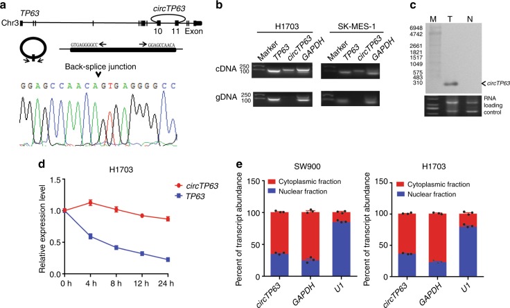
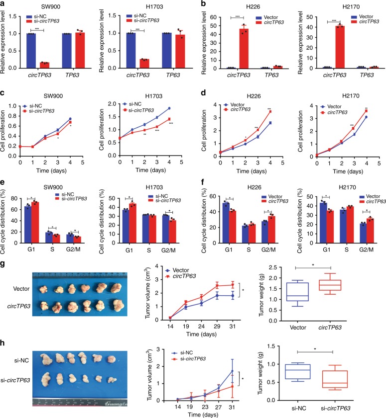
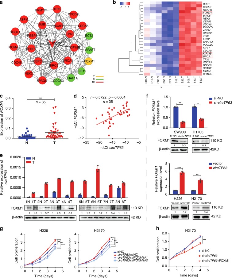
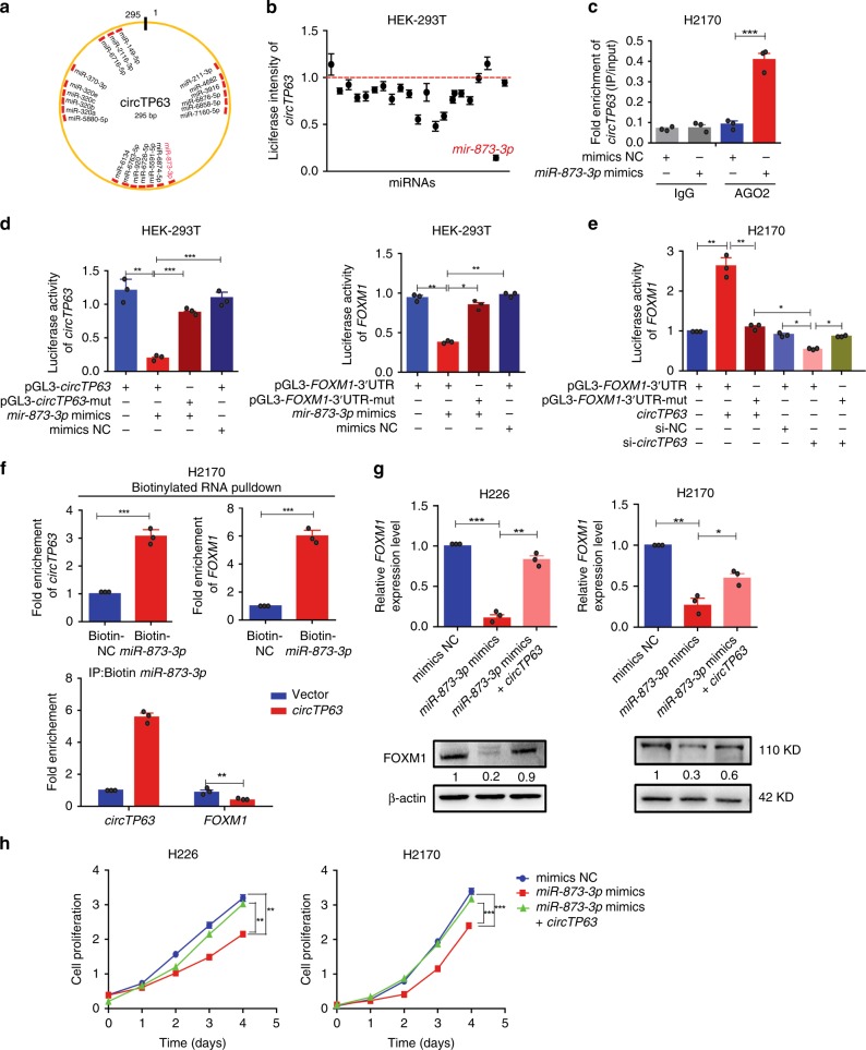
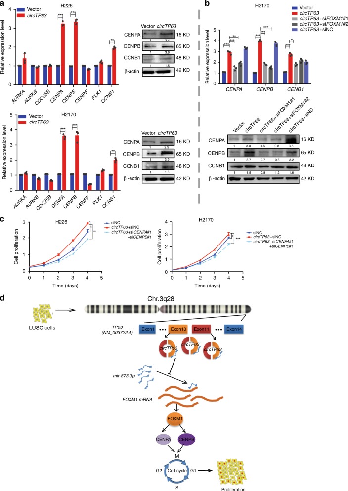
Circular RNAs (circRNAs) are identified as vital regulators in a variety of cancers. However, the role of circRNA in lung squamous cell carcinoma (LUSC) remains largely unknown. Herein, we explore the expression profiles of circRNA and mRNA in 5 paired samples of LUSC. By analyzing the co-expression network of differentially expressed circRNAs and dysregulated mRNAs, we identify that a cell cycle-related circRNA, circTP63, is upregulated in LUSC tissues and its upregulation is correlated with larger tumor size and higher TNM stage in LUSC patients. Elevated circTP63 promotes cell proliferation both in vitro and in vivo. Mechanistically, circTP63 shares miRNA response elements with FOXM1. circTP63 competitively binds to miR-873-3p and prevents miR-873-3p to decrease the level of FOXM1, which upregulates CENPA and CENPB, and finally facilitates cell cycle progression.


Conflict of interest statement

The authors declare no competing interests.

Figures

Fig. 1
Fig. 1
circRNA expression profiling reveals that circTP63 is upregulated in LUSC. a The flowchart delineates the steps for identifying and validating circRNAs in LUSC. b A heatmap shows the top 50 most upregulated and top 50 most downregulated circRNAs in five paired samples of tumorous tissues (T) and corresponding adjacent nontumorous tissues (N) from patients with LUSC by SBC Human ceRNA Array analysis. c KEGG pathway analysis for the 2832 dysregulated mRNAs. d Validation of six circRNAs by RNase R treatment and reverse transcription PCR (RT-PCR) analysis. e Fold change of circTP63 in the five paired samples of LUSC for SBC Human ceRNA Array analysis. f Left: expression levels of circTP63 in additional 35 paired samples of LUSC were determined by quantitative reverse transcription PCR (qRT-PCR). β-actin was used as a loading control. Right: histogram and pie chart of the proportions of LUSC samples in which circTP63 expression was upregulated (23/35, 65.7%, red), downregulated (7/35, 20%, blue), or no change (5/35, 14.3%, green). Log2(T/N expression) value > 1 as significantly higher expression, which <−1 as lower expression, and between −1 and 1 as no significant change. T tumorous tissue, N nontumorous tissue. The error bars (f) represent standard deviation (s.d) (n = 35). ***p< 0.001, paired t-test
Fig. 2
Fig. 2
Characterization of circTP63 in LUSC cells. a Genomic loci of circTP63 gene. circTP63 is produced at the TP63 gene (NM_003722.4) locus containing exons 10–11. The back-splice junction of circTP63 was identified by Sanger sequencing. b PCR analysis for circTP63 and its linear isoform TP63 in cDNA and genomic DNA (gDNA). c Northern blot analysis showed the size and abundance of circTP63 in one paired sample of LUSC tumorous tissue and corresponding adjacent nontumorous tissues. M: marker. d qRT-PCR for the abundance of circTP63 and TP63 in H1703 cells treated with Actinomycin D at the indicated time point. e Levels of circTP63 in the nuclear and cytoplasmic fractions of SW900 and H1703 cells. The error bars (d, e) represent s.d. (n = 3)
Fig. 3
Fig. 3
circTP63 promotes cell proliferation and tumor growth both in vitro and in vivo. a Expression levels of circTP63 and TP63 in SW900 and H1703 cells treated with circTP63 siRNA. b Expression levels of circTP63 and TP63 in H226 and H2170 cells after transduction with circTP63 lentivirus. c and d Cell proliferation analysis of LUSC cells with silencing or stably overexpressing circTP63. e and f Cell cycle analysis of LUSC cells with silencing or stably overexpressing circTP63. g The volume and weight of subcutaneous xenograft tumors of H2170 cells isolated from nude mice. h The volume and weight of subcutaneous xenograft tumors of H1703 cells isolated from nude mice; center line: median of data; Bounds of box: the second quartile to the third quartile; Whisker: minimum value to maximum value. The error bars ah represent s.d. (in af, n = 3; in g and h, n = 6). *p < 0.05; **p < 0.01; ***p < 0.001, two-tailed t-test. Source data are provided as a Source Data file
Fig. 4
Fig. 4
circTP63 contributes to cell proliferation through targeting FOXM1. a Co-expression network of circTP63 with associated 25 mRNAs. A round node represents a protein-coding gene and the arrow node represents circTP63 (hsa_circ_0068515). Lines between two nodes indicate interactions between two genes. Color represents the number of lines. b A heatmap shows mRNA levels of these 25 co-expression genes in the five paired LUSC samples of SBC Human ceRNA Array analysis. c Expression analysis for FOXM1 in additional 35 paired LUSC samples. d Correlation analysis revealed positive correlation between the levels of circTP63 and FOXM1 mRNA in the tumorous tissues of the 35 LUSC patients. ΔCt values were normalized according to β-actin. e The levels of circTP63 expression and FOXM1 protein in eight paired LUSC samples. f The mRNA and protein levels of FOXM1 in the LUSC cells with knockdown or overexpression of circTP63. g Cell proliferation assay for H226 and H2170 cells with circTP63 overexpression and FOXM1 knockdown. h Cell proliferation assay for H1703 cells with circTP63 knockdown and FOXM1 overexpression. The error bars c, eh represent s.d. (in c, n = 35; in eh, n = 3). *p < 0.05; **p < 0.01; ***p < 0.001, two-tailed t-test. Source data are provided as a Source Data file
Fig. 5
Fig. 5
circTP63 facilitates cell proliferation by relieving repression of miR-873-3p for FOXM1 expression. a A schematic model shows the putative binding sites of 22 predicted miRNAs on circTP63. b Luciferase activity of circTP63 in HEK293T cells transfected with miRNA mimics which are putative binding to the circTP63 sequence. Luciferase activity was normalized by Renila luciferase activity. c RIP was performed using AGO2 antibody in H2170 cells transfected with miR-873-3p mimics or mimics NC, then the enrichment of circTP63 was detected. d Luciferase reporter activity of circTP63 (left) and FOXM1-3′UTR (right) in HEK-293T cells co-transfected with miR-873-3p mimics or mimics NC. e Luciferase reporter activity of FOXM1–3′UTR in H2170 cells with circTP63 knockdown or overexpression. f Upper panel: circTP63 and FOXM1 were pulled down and enriched with 3′-end biotinylated miR-873-3p. Lower panel: binding activities of circTP63 and FOXM1 to 3’-end biotinylated miR-873-3p in H2170 cells with circTP63 overexpression. g FOXM1 expression in H226 and H2170 cells transfected with miR-873-3p mimics alone or co-transfected with circTP63. h Cell proliferation analysis for H226 and H2170 cells transfected with miR-873-3p mimics alone or co-transfected with circTP63. The error bars bh represent s.d. (n = 3). *p < 0.05; **p < 0.01; ***p < 0.001, two-tailed t-test. Source data are provided as a Source Data file
Fig. 6
Fig. 6
CENPA and CENPB are regulated by cicrTP63 through FOXM1. a The mRNA and protein levels of cell cycle-related genes in H226 and H2170 cells with circTP63 overexpression. b Expression changes of CENPA, CENPB, and CCNB1 after knockdown of FOXM1 in H2170 cells with circTP63 overexpression. c Cell proliferation assay for H226 and H2170 cells with circTP63 overexpression and joint knockdown of CENPA and CENPB. d Hypothesis diagram illustrates function and mechanism of circTP63 in LUSC progress. The error bars ac represent s.d. (n = 3). *p < 0.05; **p < 0.01; ***p < 0.001, two-tailed t-test. Source data are provided as a Source Data file

References

    1. Siegel RL, Miller KD, Jemal A. Cancer statistics, 2016. CA: Cancer J. Clin. 2016;66:7–30. - PubMed
    1. Chen W, et al. Cancer statistics in China, 2015. CA: Cancer J. Clin. 2016;66:115–132. - PubMed
    1. Rizvi NA, et al. Nivolumab in combination with platinum-based doublet chemotherapy for first-line treatment of advanced non-small-cell lung cancer. J. Clin. Oncol. 2016;34:2969–2979. doi: 10.1200/JCO.2016.66.9861. - DOI - PMC - PubMed
    1. Cancer Genome Atlas Research N. Comprehensive genomic characterization of squamous cell lung cancers. Nature. 2012;489:519–525. doi: 10.1038/nature11404. - DOI - PMC - PubMed
    1. Engreitz JM, et al. RNA–RNA interactions enable specific targeting of noncoding RNAs to nascent pre-mRNAs and chromatin sites. Cell. 2014;159:188–199. doi: 10.1016/j.cell.2014.08.018. - DOI - PMC - PubMed

Publication types

MeSH terms