

Pan-Renal Cell Carcinoma classification and survival prediction from histopathology images using deep learning
- PMID: 31324828
- PMCID: PMC6642160
- DOI: 10.1038/s41598-019-46718-3
Pan-Renal Cell Carcinoma classification and survival prediction from histopathology images using deep learning
Abstract
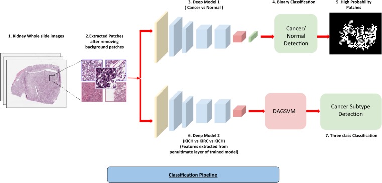
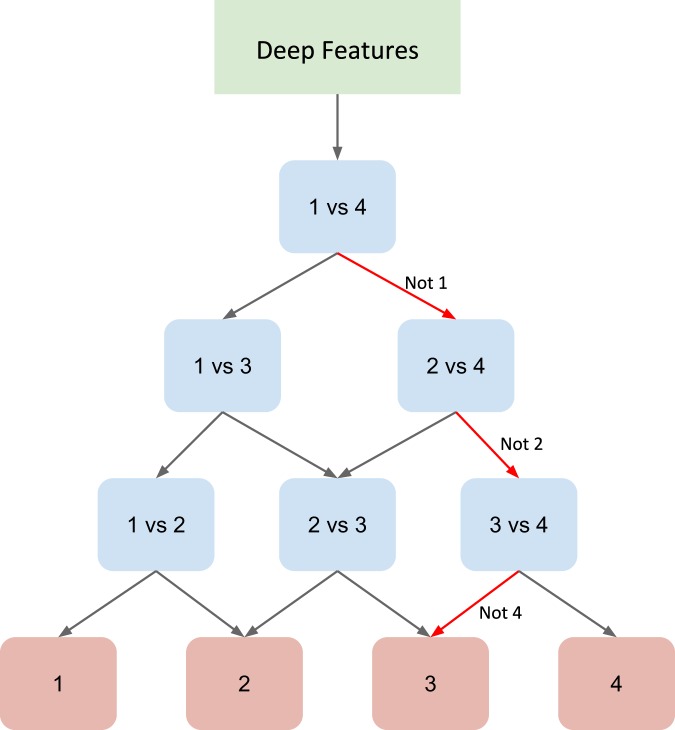
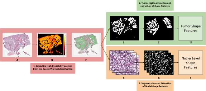
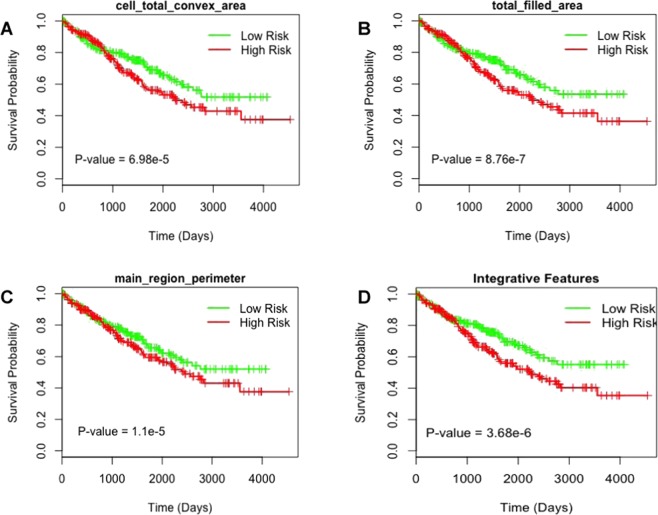
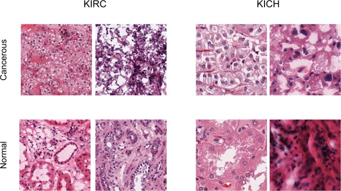
Histopathological images contain morphological markers of disease progression that have diagnostic and predictive values. In this study, we demonstrate how deep learning framework can be used for an automatic classification of Renal Cell Carcinoma (RCC) subtypes, and for identification of features that predict survival outcome from digital histopathological images. Convolutional neural networks (CNN's) trained on whole-slide images distinguish clear cell and chromophobe RCC from normal tissue with a classification accuracy of 93.39% and 87.34%, respectively. Further, a CNN trained to distinguish clear cell, chromophobe and papillary RCC achieves a classification accuracy of 94.07%. Here, we introduced a novel support vector machine-based method that helped to break the multi-class classification task into multiple binary classification tasks which not only improved the performance of the model but also helped to deal with data imbalance. Finally, we extracted the morphological features from high probability tumor regions identified by the CNN to predict patient survival outcome of most common clear cell RCC. The generated risk index based on both tumor shape and nuclei features are significantly associated with patient survival outcome. These results highlight that deep learning can play a role in both cancer diagnosis and prognosis.
Conflict of interest statement
The authors declare no competing interests.
Figures
References
-
- American cancer society, http://www.cancer.org/cancer/kidneycancer. Accessed: 2018-10-1.
-
- Kovacs G, et al. The heidelberg classification of renal cell tumours. The J. Pathol. A J. Pathol. Soc. Gt. Br. Irel. 1997;183:131–133. - PubMed
-
- Truong LD, Shen SS. Immunohistochemical diagnosis of renal neoplasms. Arch. pathology & laboratory medicine. 2011;135:92–109. - PubMed
Publication types
MeSH terms
LinkOut - more resources
Full Text Sources
Other Literature Sources
Medical
