Comparative Study
. 2019 Jul 19;9(1):10508.
doi: 10.1038/s41598-019-46606-w.

A direct comparison of interphase FISH versus low-coverage single cell sequencing to detect aneuploidy reveals respective strengths and weaknesses

Affiliations
Comparative Study

A direct comparison of interphase FISH versus low-coverage single cell sequencing to detect aneuploidy reveals respective strengths and weaknesses

Grasiella A Andriani et al. Sci Rep. .

Abstract

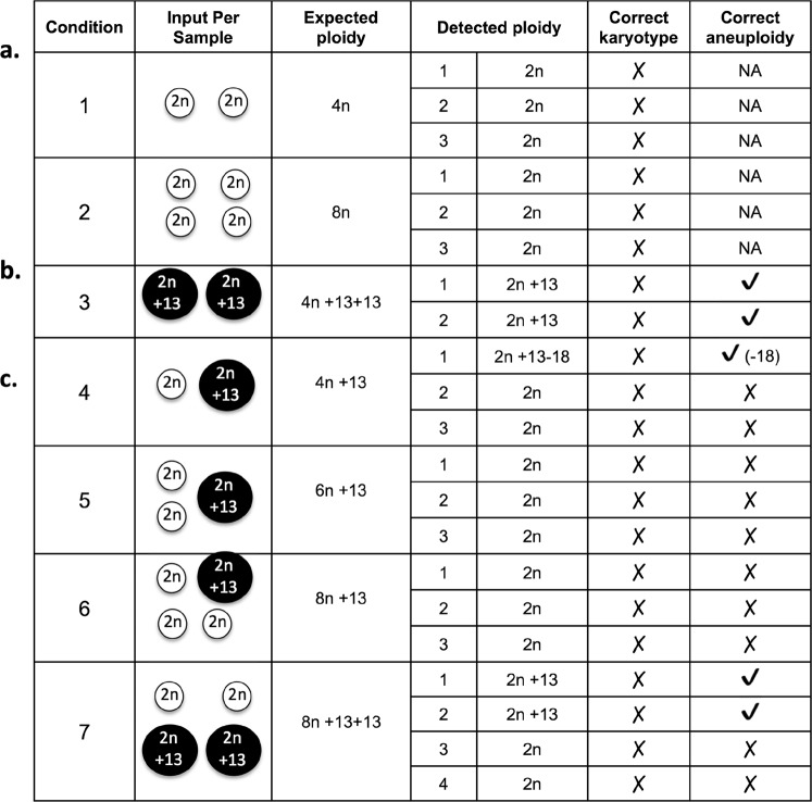
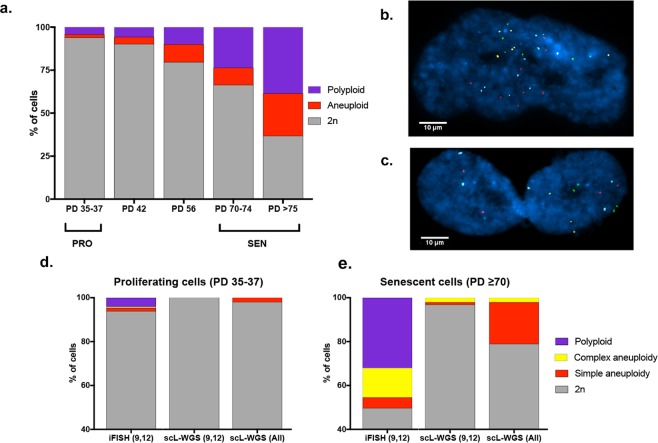
Aneuploidy has been reported to occur at remarkably high levels in normal somatic tissues using Fluorescence In Situ Hybridization (FISH). Recently, these reports were contradicted by single-cell low-coverage whole genome sequencing (scL-WGS) analyses, which showed aneuploidy frequencies at least an order of magnitude lower. To explain these seemingly contradictory findings, we used both techniques to analyze artificially generated mock aneuploid cells and cells with natural random aneuploidy. Our data indicate that while FISH tended to over-report aneuploidies, a modified 2-probe approach can accurately detect low levels of aneuploidy. Further, scL-WGS tends to underestimate aneuploidy levels, especially in a polyploid background.


Conflict of interest statement

The authors declare no competing interests.

Figures

Figure 1
Figure 1
Mock aneuploidy and aneuploidy detected by scL-WGS. (a) Polyploidy, generated by combining two or four 2n cells into a tube prior to WGA. (b,c) Aneuploidy in a polyploid background, generated by combining 2n and T13 cells, as illustrated. The detected ploidy and aneuploidy are indicated to the right where detection of the correct karyotype and/or aneuploidy is also shown.
Figure 2
Figure 2
Ploidy analysis of IMR-90 human primary fibroblasts by iFISH and scL-WGS. (a) Ploidy distribution at different population doublings (PDs) of IMR-90 cells, measured by 4-color iFISH. Proliferating cells (PRO, N = 605) were analyzed at population doubling levels (PDL) 35–37 and replicatively senescent cells (SEN, N = 905) analyzed from PDL 70 and beyond. (b,c) Representative images of a cell (b) with complex aneuploidy and (c) binucleated tetraploid cell. (d) Percentage of non-diploid (Not 2n) cells detected by iFISH and scL-WGS in PRO (N = 51) and (e) SEN (N = 95) cells. scL-WGS (All) refers to the levels of Not 2n cells considering all chromosomes. scL-WGS (9,12) refers to the levels of Not 2n cells considering only chromosomes 9 and 12, which were also analyzed by iFISH.

References

    1. Rehen SK, et al. Constitutional aneuploidy in the normal human brain. The Journal of neuroscience: the official journal of the Society for Neuroscience. 2005;25:2176–2180. doi: 10.1523/JNEUROSCI.4560-04.2005. - DOI - PMC - PubMed
    1. Rehen SK, et al. Chromosomal variation in neurons of the developing and adult mammalian nervous system. Proceedings of the National Academy of Sciences of the United States of America. 2001;98:13361–13366. doi: 10.1073/pnas.231487398. - DOI - PMC - PubMed
    1. Faggioli F, Wang T, Vijg J, Montagna C. Chromosome-specific accumulation of aneuploidy in the aging mouse brain. Human molecular genetics. 2012;21:5246–5253. doi: 10.1093/hmg/dds375. - DOI - PMC - PubMed
    1. Yurov YB, et al. The variation of aneuploidy frequency in the developing and adult human brain revealed by an interphase FISH study. J Histochem Cytochem. 2005;53:385–390. doi: 10.1369/jhc.4A6430.2005. - DOI - PubMed
    1. Andriani GA, et al. Whole chromosome aneuploidy in the brain of Bub1bH/H and Ercc1-/Delta7 mice. Human molecular genetics. 2016;25:755–765. doi: 10.1093/hmg/ddv612. - DOI - PMC - PubMed

Publication types