. 2019 Jul 19;9(1):10475.
doi: 10.1038/s41598-019-47006-w.

A zebrafish drug screening platform boosts the discovery of novel therapeutics for spinal cord injury in mammals

Affiliations

A zebrafish drug screening platform boosts the discovery of novel therapeutics for spinal cord injury in mammals

Diana Chapela et al. Sci Rep. .

Erratum in

Abstract

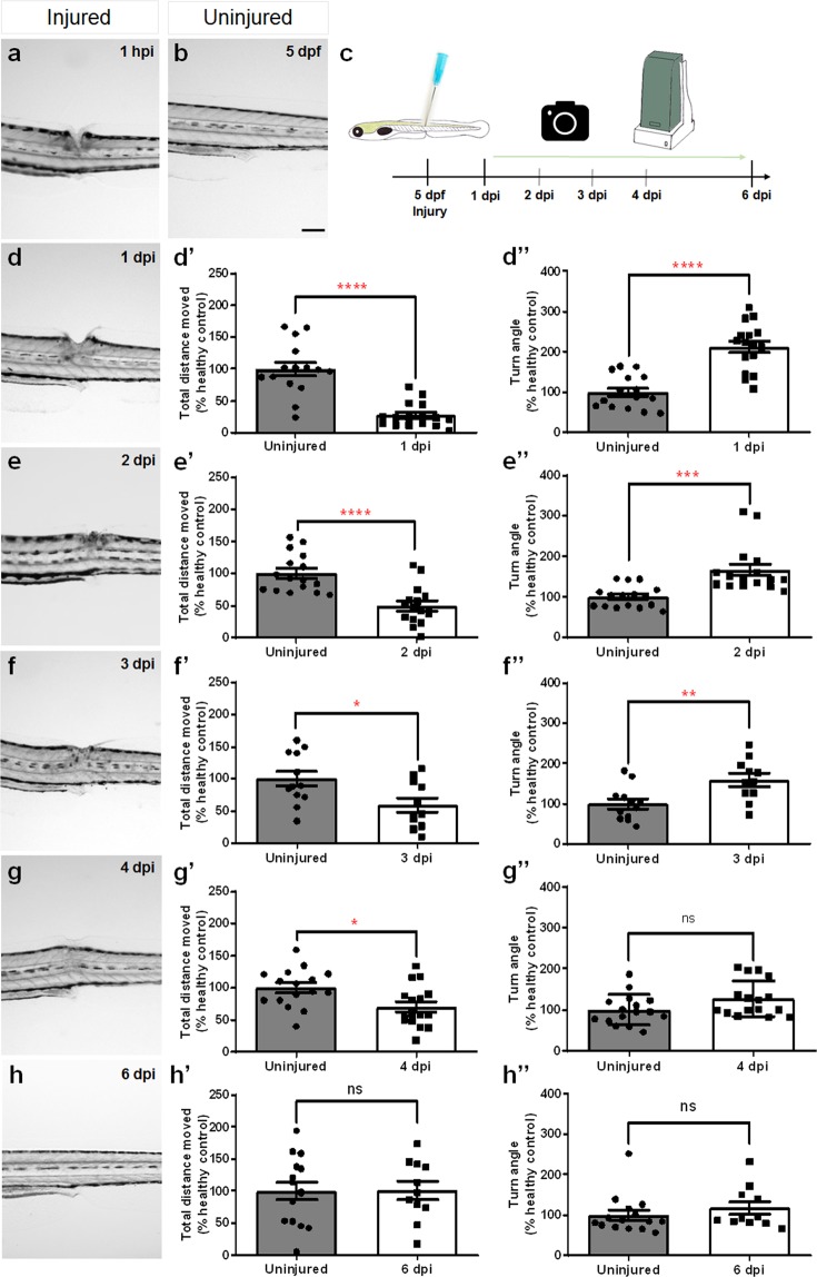
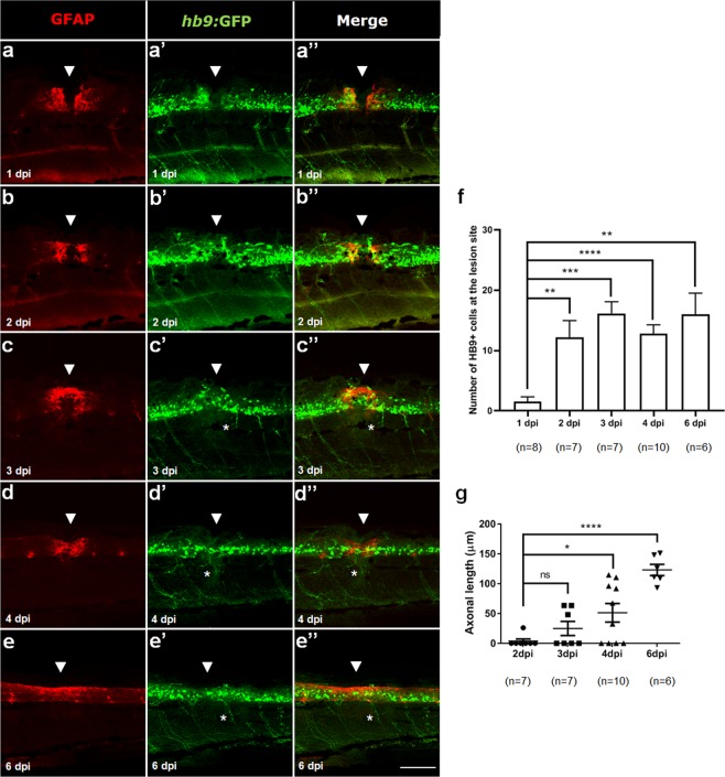
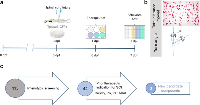
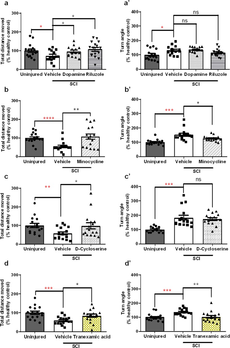
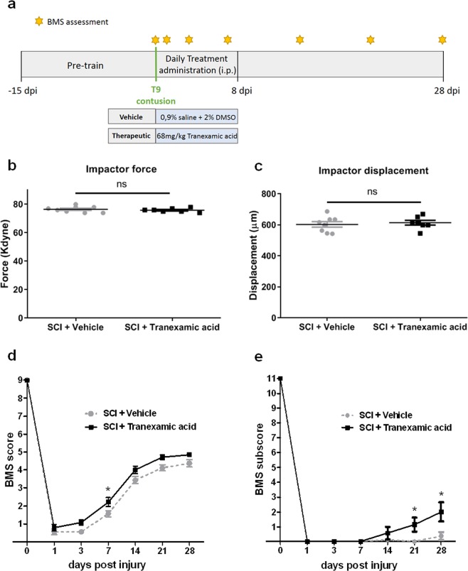
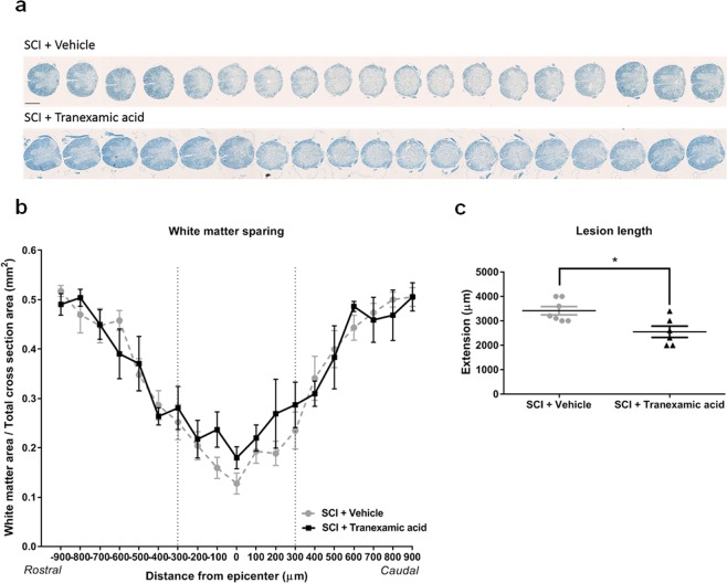
Spinal cord injury (SCI) is a complex condition, with limited therapeutic options, that results in sensory and motor disabilities. To boost discovery of novel therapeutics, we designed a simple and efficient drug screening platform. This innovative approach allows to determine locomotor rescue properties of small molecules in a zebrafish (Danio rerio) larval spinal cord transection model. We validated our screening platform by showing that Riluzole and Minocycline, two molecules that are in clinical trials for SCI, promote rescue of the locomotor function of the transected larvae. Further validation of the platform was obtained through the blind identification of D-Cycloserine, a molecule scheduled to enter phase IV clinical trials for SCI. Importantly, we identified Tranexamic acid and further showed that this molecule maintains its locomotor recovery properties in a rodent female contusion model. Our screening platform, combined with drug repurposing, promises to propel the rapid translation of novel therapeutics to improve SCI recovery in humans.


Conflict of interest statement

The authors declare no competing interests.

Figures

Figure 1
Figure 1
Temporal profile of tissue regeneration and locomotor recovery upon spinal cord transection in 5 dpf zebrafish larvae. (b) Brightfield image of the trunk/tail area of an injured 5 dpf larva. (c) Experimental design for the analysis of the temporal profile of tissue regeneration and locomotor recovery. (a,dh) Brightfield images of the recovery of zebrafish larvae with a lesion in the dorsal trunk area from 1 hpi to 6 dpi. (d’h’) Total distance moved and (d”h”) turn angle parameters of injured larvae and age-matched uninjured controls at 1 dpi (n = 16 uninjured larvae, n = 16 injured larvae), 2 dpi (n = 16 uninjured larvae, n = 16 injured larvae), 3 dpi (n = 12 uninjured larvae, n = 11 injured larvae), 4 dpi (n = 16 uninjured larvae, n = 16 injured larvae) and 6 dpi (n = 15 uninjured larvae, n = 11 injured larvae). dpf_days-post-fertilization, hpi_hours-post-injury. Mean ± s.e.m. of one experiment is presented. ns - not significant, *p < 0.05, **p < 0.01, ***p < 0.001 and ****p < 0.0001, Student’s t-test with Welch’s correction. Scale bar: 200 µm.
Figure 2
Figure 2
Glial bridge formation and motor neurons regeneration after SCI in 5 dpf larvae. Labeling pattern of Hb9:GFP transgenic larvae with double immunohistochemistry against GFAP (red) to reveal the glial bridge (ae) and against GFP (green) to reveal HB9+ motor neurons (a’e’) from 1 to 6 dpi. (a”e”) Merged channels for each time point. White arrowheads show the lesion site and asterisks highlight the regenerating HB9+ peripheral axons. Rostral side is to the left and dorsal side is up. Number of HB9+ motor neurons (f) and length of HB9+ peripheral axons (g) at the lesion site from 1 to 6 dpi. SCI_spinal cord injury, dpf_days-post-fertilization, hpi_hours-post-injury Mean ± s.e.m. of one experiment is presented. ns - not significant, *p < 0.05, **p < 0.01, ***p < 0.001 and ****p < 0.0001, Student’s t-test with Welch’s correction. Scale bar: 100 µm.
Figure 3
Figure 3
Schematic representation of the platform developed for the selection of novel compounds with SCI recovery properties. (a) The protocol of the phenotypic screening starts with the spinal cord transection of 5 days-post-fertilization (dpf) Hb9:GFP transgenic zebrafish larvae. The next day (1 dpi), the transected larvae are transferred into a 6-well plate. The therapeutics are added to the medium and zebrafish larvae are incubated for 24 hours. At 2 dpi, the behavioural analysis is conducted in a 96-well plate with a video tracking system. (b) Total distance moved and turn angle are used as indicators of locomotor function. (c) Schematic representation of the sequential criteria steps that led to the identification of the 3 potential new therapeutics for SCI.
Figure 4
Figure 4
Rescue of motor impairments by different pharmaceutical classes of molecules in a transected zebrafish larval model of SCI. Locomotor performance depicted from total distance moved (a,b) and turn angle (a’,b’) in transected zebrafish larvae treated with 30 µM of Dopamine (n = 14 larvae), 0,5 µM of Riluzole (n = 16 larvae) and 25 µM of Minocycline (n = 16 larvae) as compared to vehicle treated injured larvae (n = 13–15 larvae). Locomotor performance depicted from total distance moved (c,d) and turn angle (c’,d’) in transected zebrafish larvae treated with D-Cycloserine (n = 16 larvae) and Tranexamic acid (n = 16 larvae), both picked from the library of FDA approved chemical compounds, as compared to vehicle treated injured larvae (n = 16 larvae). All percentages are relative to the mean of vehicle treated uninjured larvae (healthy control, n = 16 larvae). Mean ± s.e.m. of four independent experiments is presented. ns - not significant, *p < 0.05, **p < 0.01, ***p < 0.001 and ****p < 0.0001, Student’s t-test with Welch’s correction.
Figure 5
Figure 5
Locomotor recovery after T9 contusion in a rodent SCI model. (a) Experimental design of the Tranexamic acid efficacy test in a T9 contusion rodent model of SCI. (b) Impact force (kilodynes) imparted on the spinal cord during SCI (ns - not significant, t student test with Welch’s correction). (c) Displacement (μm) of the impactor tip upon contact with the spinal cord during T9 contusion (ns - not significant, t student test with Welch’s correction). (d) BMS scores of Tranexamic acid-treated mice compared with vehicle-treated mice from 1 to 28 dpi. (e) BMS subscores of Tranexamic acid-treated mice compared with the vehicle-treated mice from 1 to 28 dpi. Mean ± s.e.m. of one experiment is presented. *p < 0.05, Two-way repeated measures ANOVA followed by Bonferroni’s post-hoc correction. n = 8 for SCI+ Vehicle and n = 7 for SCI+ Tranexamic acid.
Figure 6
Figure 6
White matter sparing and lesion length in vehicle-treated mice and Tranexamic acid-treated mice at 28 days after SCI. (a) Representative spinal cord sections of vehicle-treated mice comparing to Tranexamic acid- treated mice labelled with luxol fast blue staining. Scale bar: 500 µm. (b) The white matter area per total cross section area from the epicenter to 900 µm on the rostral and caudal sides in vehicle-treated mice and in Tranexamic acid-treated mice. Two-way ANOVA followed by Bonferroni post hoc correction. n = 7 for SCI+ Vehicle and n = 6 for SCI+ Tranexamic acid. (c) Lesion length in vehicle-treated mice (n = 7) and in Tranexamic acid-treated mice (n = 6). *p < 0.05, Student’s t-test with Welch’s correction.

References

    1. Yılmaz T, Kaptanoğlu E. Current and future medical therapeutic strategies for the functional repair of spinal cord injury. World J Orthop. 2015;6:42–55. doi: 10.5312/wjo.v6.i1.42. - DOI - PMC - PubMed
    1. Duncan GJ, et al. Locomotor recovery following contusive spinal cord injury does not require oligodendrocyte remyelination. Nat Commun. 2018;9:3066. doi: 10.1038/s41467-018-05473-1. - DOI - PMC - PubMed
    1. Oudega M. Molecular and cellular mechanisms underlying the role of blood vessels in spinal cord injury and repair. Cell Tissue Res. 2012;349:269–288. doi: 10.1007/s00441-012-1440-6. - DOI - PubMed
    1. Boutonnet M, Laemmel E, Vicaut E, Duranteau J, Soubeyrand M. Combinatorial therapy with two pro-coagulants and one osmotic agent reduces the extent of the lesion in the acute phase of spinal cord injury in the rat. Intensive Care Med Exp. 2017;5:51. doi: 10.1186/s40635-017-0164-z. - DOI - PMC - PubMed
    1. Zhou X, He X, Ren Y. Function of microglia and macrophages in secondary damage after spinal cord injury. Neural Regen Res. 2014;9:1787–1795. doi: 10.4103/1673-5374.143423. - DOI - PMC - PubMed

Publication types