. 2019 Oct:81:545-559.
doi: 10.1016/j.bbi.2019.07.015. Epub 2019 Jul 17.

A permethrin metabolite is associated with adaptive immune responses in Gulf War Illness

Affiliations

A permethrin metabolite is associated with adaptive immune responses in Gulf War Illness

Utsav Joshi et al. Brain Behav Immun. 2019 Oct.

Abstract

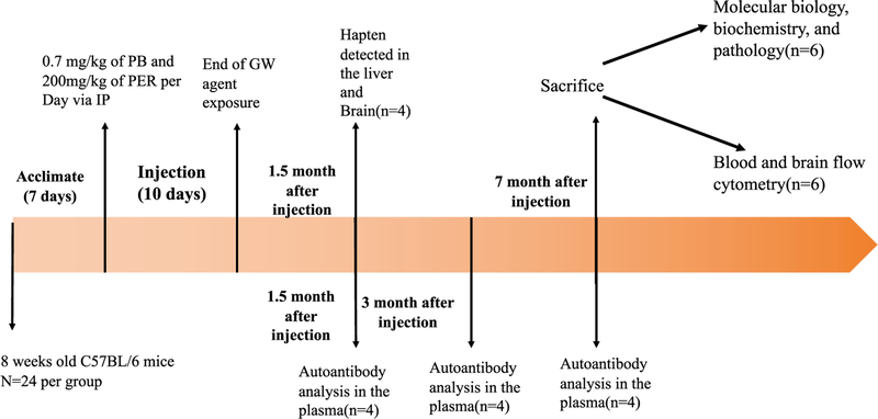
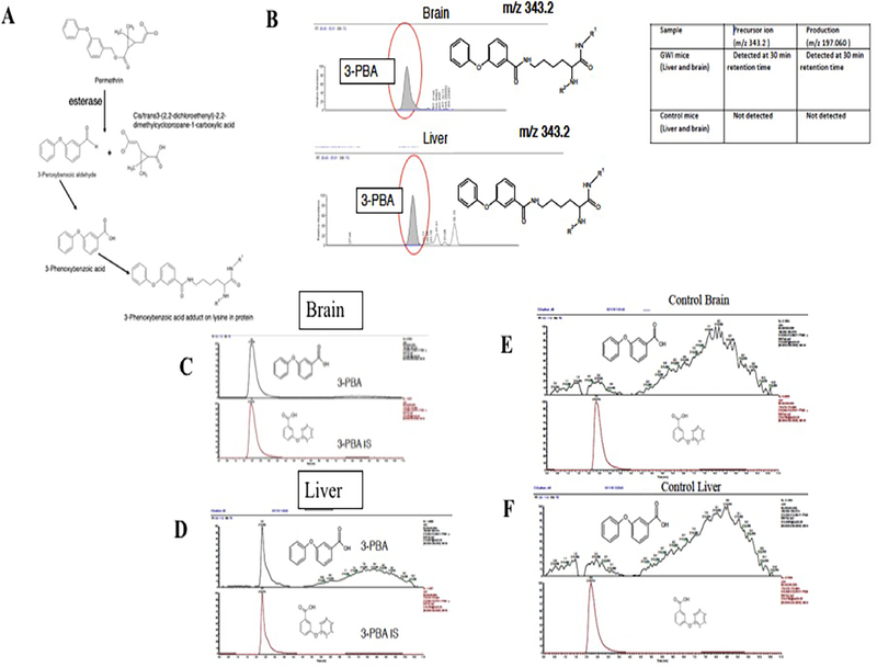
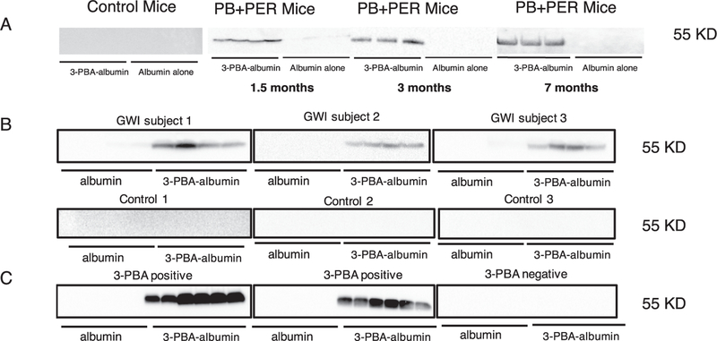
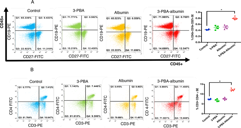
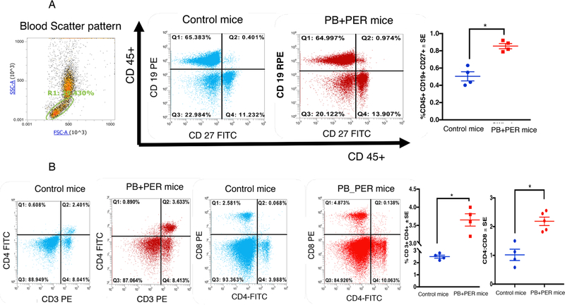
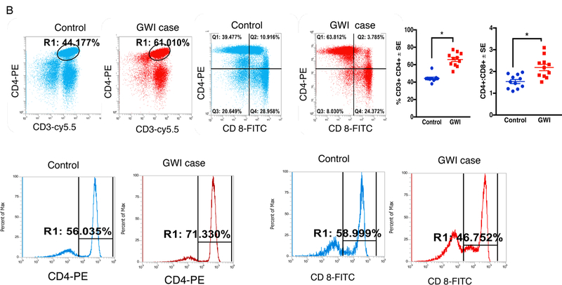
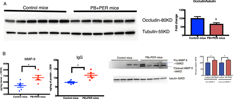
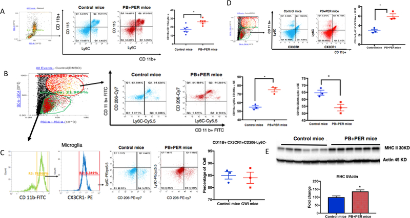
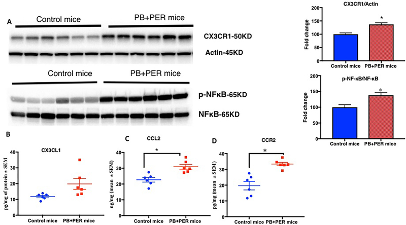
Gulf War Illness (GWI), affecting 30% of veterans from the 1991 Gulf War (GW), is a multi-symptom illness with features similar to those of patients with autoimmune diseases. The objective of the current work is to determine if exposure to GW-related pesticides, such as permethrin (PER), activates peripheral and central nervous system (CNS) adaptive immune responses. In the current study, we focused on a PER metabolite, 3-phenoxybenzoic acid (3-PBA), as this is a common metabolite previously shown to form adducts with endogenous proteins. We observed the presence of 3-PBA and 3-PBA modified lysine of protein peptides in the brain, blood and liver of pyridostigmine bromide (PB) and PER (PB+PER) exposed mice at acute and chronic post-exposure timepoints. We tested whether 3-PBA-haptenated albumin (3-PBA-albumin) can activate immune cells since it is known that chemically haptenated proteins can stimulate immune responses. We detected autoantibodies against 3-PBA-albumin in plasma from PB + PER exposed mice and veterans with GWI at chronic post-exposure timepoints. We also observed that in vitro treatment of blood with 3-PBA-albumin resulted in the activation of B- and T-helper lymphocytes and that these immune cells were also increased in blood of PB + PER exposed mice and veterans with GWI. These immune changes corresponded with elevated levels of infiltrating monocytes in the brain and blood of PB + PER exposed mice which coincided with alterations in the markers of blood-brain barrier disruption, brain macrophages and neuroinflammation. These studies suggest that pesticide exposure associated with GWI may have resulted in the activation of the peripheral and CNS adaptive immune responses, possibly contributing to an autoimmune-type phenotype in veterans with GWI.

Keywords: Autoantibody; Gulf War Illness; Hapten; Permethrin; Pesticide.


Conflict of interest statement

Declaration of Competing Interest

The author(s) declares no competing interests. The contents do not represent the views of the Department of Veterans Affairs or the United States Government.

Figures

Fig. 1.
Fig. 1.
Timeline of study design.
Fig. 2.
Fig. 2.
Metabolites of PER pesticide is detected as protein adducts in the brain and liver homogenates. (A) A schematic of PER metabolites. (B) Detection of precursor and product ion of 3-PBA lysine (343.2) and its product ions (197.06) in the brain and liver homogenates from a GWI mouse sample. (C and D) Using LC-MS and LC-MS/MS, we detected 3-PBA (m/z 213.055 at 3 min) and its PRM product ion (m/z 213.1 > 93.033 at that same time) in the brain and liver (mean ± SE (n = 4 or 5/ group). While only protein-conjugated 3-PBA recovered after hydrolysis was detected in the brain, free 3-PBA and protein-conjugated 3-PBA were detected in the liver at 1.5 months post-exposure. (E and F) Peak plots show absence of 3-PBA (m/z 213.055 at 3 min) and its transition ion (m/z 93.033) ions in control mice. These peak plots show presence of 3-PBA internal standard 219.1 at 3 min and its transition ion (m/z 93.033) ions in control mice.
Fig. 3.
Fig. 3.
Chronic elevation of autoantibodies against 3-PBA-albumin in plasma of PB + PER mice, in veterans with GWI and in farm workers exposed to pyrethroids. Western Blot showed that control mice had no cross-reactivity to 3-PBA-albumin or albumin alone, but cross-reactivity in PB + PER mice plasma was observed for 3-PBA-albumin at 1.5-, 3- and 7-months post-exposure in PB + PER mice (n = 4 or 5/group). (B) Autoantibodies against 3- PBA-albumin were detected in 12 out of 14 veterans with GWI and 1 out of 10 control GW veterans. (C) Immunoreactivity of plasma with 3- PBA-albumin was observed in 5 out of 5 pyrethroid-exposed farm workers or ate pyrethroid tainted fruits and vegetables (as confirmed using 3-PBA measurements in urine), and a lack of immunoreactivity in controls in whom urine 3-PBA was undetected.
Fig. 4.
Fig. 4.
3-PBA-albumin treatment increased memory B-cell and T-helper cells in 7 months old control mice. Mean ± SE (n = 4 per group). (A) After overnight incubation of murine blood with 3-PBA, albumin and 3-PBA-albumin, antigen-responsive CD45+ CD19+CD27+ B-cells are significantly elevated in 3-PBA-albumin compared to all other treatments. (B) T-helper cells, CD3+CD4+ , were also elevated upon 3-PBA-albumin treatment. Comparisons between 3-PBA and controls and albumin and controls were not significant. *p < 0.05.
Fig. 5.
Fig. 5.
Antigen-activated B- and T-cells are increased in blood of PB + PER mice at 7 months post-exposure. Mean ± SE (n = 4/5 per group). (A) Increased staining of antigen-responsive CD19+ CD27+ B-cells were detected in GWI compared to control mice. (B) Panel B shows that CD3+CD4+ Txells are increased in blood of PB + PER mice. However, CD8+ T.cells did not differ between the two groups. Ratios of CD4:CD8 was significantly elevated in PB + PER mice. *p < 0.05.
Fig. 6.
Fig. 6.
Memory B- and CD4 T-cells are increased in blood of veterans with GWI. Mean ± SE (n = 12 per group). (A) Increased staining CD19+ and CD19+ CD27 + memory B-cells in blood of veterans with GWI compared to healthy GW control veterans. (B) CD3+CD4+ TH cells are increased in blood of veterans with GWI. However, CD8+ TH cells did not significantly differ between control and GWI veterans. However, there were no group differences for CD3+CD8+ TH cells. *p < 0.05.
Fig. 6.
Fig. 6.
Memory B- and CD4 T-cells are increased in blood of veterans with GWI. Mean ± SE (n = 12 per group). (A) Increased staining CD19+ and CD19+ CD27 + memory B-cells in blood of veterans with GWI compared to healthy GW control veterans. (B) CD3+CD4+ TH cells are increased in blood of veterans with GWI. However, CD8+ TH cells did not significantly differ between control and GWI veterans. However, there were no group differences for CD3+CD8+ TH cells. *p < 0.05.
Fig. 7.
Fig. 7.
PB + PER mice showed evidence of BBB impairment. Mean ± SE (n = 6 per group). (A) Occludin was decreased in the brains of PB + PER mice whereas (B) MMP-9 and IgG were increased in PB + PER mice brain. (C) Representative images of MMP-9 western blot of whole brain homogenate. Band intensities from control and PB + PER animal brain tissue (n = 4). *p < 0.05.
Fig. 8.
Fig. 8.
Infiltrating monocytes are increased in blood and brains of PB + PER mice at 7–8 months post-exposure. Mean ± SE (n=12 per group) (A) Increases in blood monocyte gated by CD11B+ CD115+ and Ly6C+ in PB + PER mice were observed. (B) PB + PER mice brain showed increased macrophage population as detected by CD11b+ CD 206+ Ly6C+ but (C) no change in microglia population detected by CD11b+ CX3CR1+ and CD206-. (D) Infiltrating monocytes converting to macrophage (CD11b+ , CX3CR1− and Ly6C+) were also increased in the brains of PB + PER mice. (E) Increase in MHC-II in the brain of PB + PER mice was detected by Western Blot *p < 0.05.
Fig. 9.
Fig. 9.
CCR2 and its ligand CCL2 are chronically increased in the brains of PB + PER-exposed mice leading to inflammation. Mean as % control ± SEM (n = 6 per group). (A) Western Blot showing CX3CR1/actin and pNFκB/NFκB is increased in the brains of PB + PER-exposed mice. (B) No change in CX3CL1 level as measured by ELISA. (C) Levels of CCL2 were also increased in PB + PER-exposed mice at 7-months post-exposure. (D) CCR2 levels were increased in the brains of PB + PER mice. *p < 0.05.

References

    1. Abdel-Rahman A, et al., 2004. Stress and combined exposure to low doses of pyridostigmine bromide, DEET, and permethrin produce neurochemical and neuropathological alterations in cerebral cortex, hippocampus, and cerebellum. J. Toxicol. Environ. Health, Part A 67 (2), 163–192. 10.1080/15287390490264802. - DOI - PubMed
    1. Abdullah L, et al., 2011. Proteomic CNS profile of delayed cognitive impairment in mice exposed to Gulf War agents. NeuroMol. Med. 13 (4), 275–288. 10.1007/s12017-011-8160-z. - DOI - PubMed
    1. Abdullah L, et al., 2012. Lipidomic profiling of phosphocholine containing brain lipids in mice with sensorimotor deficits and anxiety-like features after exposure to gulf war agents. NeuroMol. Med. 14 (4), 349–361. 10.1007/s12017-012-8192-z. - DOI - PubMed
    1. Abdullah L, et al., 2016. Translational potential of long-term decreases in mitochondrial lipids in a mouse model of Gulf War Illness. Toxicology 372, 22–33. 10.1016/j.tox.2016.10.012. Elsevier Ireland Ltd. - DOI - PubMed
    1. Abou-Donia MB, et al., 2017. Screening for novel central nervous system biomarkers in veterans with Gulf War Illness. Neurotoxicol. Teratol. 61, 36–46. 10.1016/j.ntt.2017.03.002. - DOI - PubMed

Publication types

MeSH terms