Review
. 2019 Oct 1:461:123-131.
doi: 10.1016/j.canlet.2019.07.012. Epub 2019 Jul 18.

Notch signaling in breast cancer: From pathway analysis to therapy

Affiliations
Review

Notch signaling in breast cancer: From pathway analysis to therapy

B Madhu Krishna et al. Cancer Lett. .

Abstract

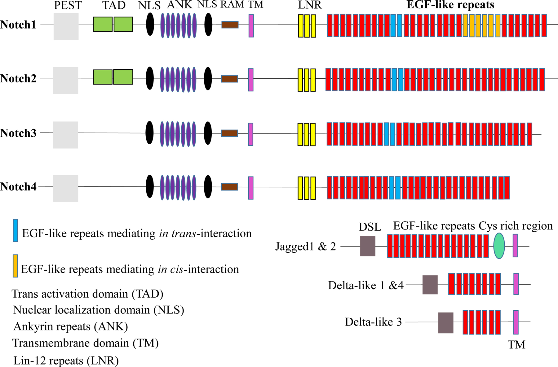
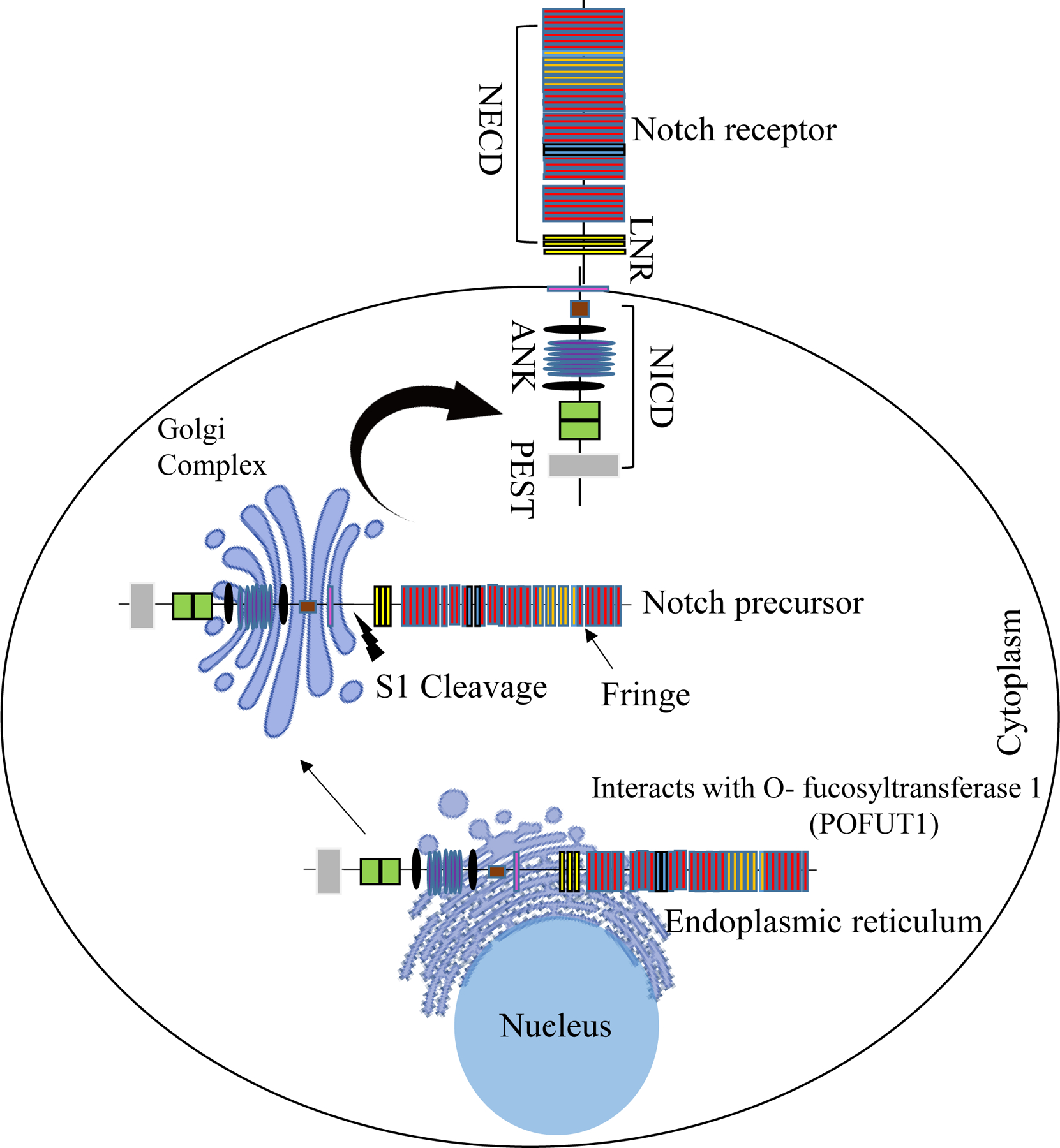
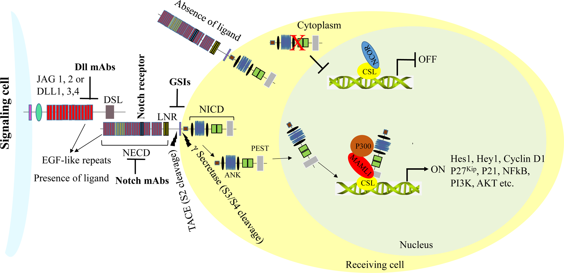
The Notch signaling pathway, which is highly conserved from sea urchins to humans, plays an important role in cell-differentiation, survival, proliferation, stem-cell renewal, and determining cell fate during development and morphogenesis. It is well established that signaling pathways are dysregulated in a wide-range of diseases, including human malignancies. Studies suggest that the dysregulation of the Notch pathway contributes to carcinogenesis, cancer stem cell renewal, angiogenesis, and chemo-resistance. Elevated levels of Notch receptors and ligands have been associated with cancer-progression and poor survival. Furthermore, the Notch signaling pathway regulates the transcriptional activity of key target genes through crosstalk with several other signaling pathways. Indeed, increasing evidence suggests that the Notch signaling pathway may serve as a therapeutic target for the treatment of several cancers, including breast cancer. Researchers have demonstrated the anti-tumor properties of Notch inhibitors in various cancer types. Currently, Notch inhibitors are being evaluated for anticancer efficacy in a number of clinical-trials. However, because there are multiple Notch receptors that can exhibit either oncogenic or tumor-suppressing roles in various cells, it is important that the Notch inhibitors are specific to particular receptors that are tumorigenic in nature. This review critically evaluates existing Notch inhibitory drugs and strategies and summarizes the previous discoveries, current understandings, and recent developments in support of Notch receptors as therapeutic targets in breast cancer.

Keywords: Breast cancer; Chemo-resistance; Monoclonal antibodies; Notch receptor; γ-secretase inhibitors.


Conflict of interest statement

Conflict of interest: The authors have no conflicts of interest to disclose.

Figures

Figure 1:
Figure 1:. Structure of Notch receptors and ligands
Notch proteins are a highly conserved family of transmembrane receptors. Notch receptors and ligands contain multiple domains. The extracellular domains (NECDs) of Notch receptors 1–4 and their ligands (Jagged 1, Jagged 2, Dll1, Dll3, and Dll4) contain EGF-like repeats. Notch 1 and Notch 2 contain 36 EGF-like repeats, whereas Notch 3 and Notch 4 contain 34 and 29, respectively. The intracellular domains (NICDs) of Notch 1 and 2 contains a RAM (RBP-jk association molecule) domain, NLSs (Nuclear localization signals), an ANK (Ankyrin repeat) domain, a TAD (Trans-activation domain), and a PEST domain. The NICDs of Notch 3 and Notch 4 are similar, but the TAD is absent in both. The extracellular domain of Serrate-like ligands Jagged 1 and Jagged 2 consists of a DSL domain, EGF-like repeats, and a Cys-rich region. The extracellular domain of the Delta-like ligands (Dll1, Dll3, and Dll4) is similar, but the Cys-rich region is absent.
Figure 2:
Figure 2:. Maturation of Notch receptors
Notch receptors mature in the endoplasmic reticulum (ER) and Golgi complex. Fucosylation is essential and occurs through the interaction between the Notch precursor protein and O-fucosyltransferase 1 (OFUT1 in Drosophila, POFUT1 in mammals) in the ER. The fucosylated Notch precursor is then transported to the Golgi complex, where proteolytic cleavage by Furin-like convertase at site 1 (S1) occurs. Finally, the matured Notch is transported to the cell surface.
Figure 3:
Figure 3:. Schematic representation of Notch receptor activation
Notch receptors are activated upon binding to Serrate- and Delta-like ligands present on the cell membranes of adjacent cells. Following successful activation, Notch receptors undergo a series of proteolytic cleavages at site 2 (S2), mediated by metalloprotease 10 (ADAM10) and TACE (TNFα converting enzyme). Additional proteolytic cleavages at the transmembrane domain (NTD) are carried out by a multi-subunit complex, γ-secretase, at site 3 (S3). The Notch intracellular domains (NICDs) are then released into the cytoplasm. The NICDs further translocate into the nucleus, where they displace histone deacetylase and co-repressors in CSL repressor complexes and recruit MAML1 and histone acetyltransferase p300 to form active transcriptional complexes, which regulate the transcriptional activity of Notch target genes.

References

    1. Theodosiou A, Arhondakis S, Baumann M, Kossida S. Evolutionary scenarios of Notch proteins. Mol Biol Evol 2009; 26: 1631–40. - PubMed
    1. Xu J, Gridley T. Notch Signaling during Oogenesis in Drosophila melanogaster. Gene Res Intl 2012; 2012: 648207. - PMC - PubMed
    1. Rebay I, Fleming RJ, Fehon RG, Cherbas L, Cherbas P, Artavanis-Tsakonas S. Specific EGF repeats of Notch mediate interactions with Delta and Serrate: implications for Notch as a multifunctional receptor. Cell 1991; 67: 687–99. - PubMed
    1. Weinmaster G, Roberts VJ, Lemke G. Notch2: a second mammalian Notch gene. Development 1992; 116: 931–41. - PubMed
    1. Ran Y, Hossain F, Pannuti A, Lessard CB, Ladd GZ, Jung JI, Minter LM, Osborne BA, Miele L, Golde TE. Gamma-secretase inhibitors in cancer clinical trials are pharmacologically and functionally distinct. EMBO Molecular Medicine 2017; 9: 950–66. - PMC - PubMed

Publication types

MeSH terms