. 2019 Aug 19;29(16):2665-2675.e4.
doi: 10.1016/j.cub.2019.06.082. Epub 2019 Jul 18.

A Single MicroRNA-Hox Gene Module Controls Equivalent Movements in Biomechanically Distinct Forms of Drosophila

Affiliations

A Single MicroRNA-Hox Gene Module Controls Equivalent Movements in Biomechanically Distinct Forms of Drosophila

A Raouf Issa et al. Curr Biol. .

Abstract

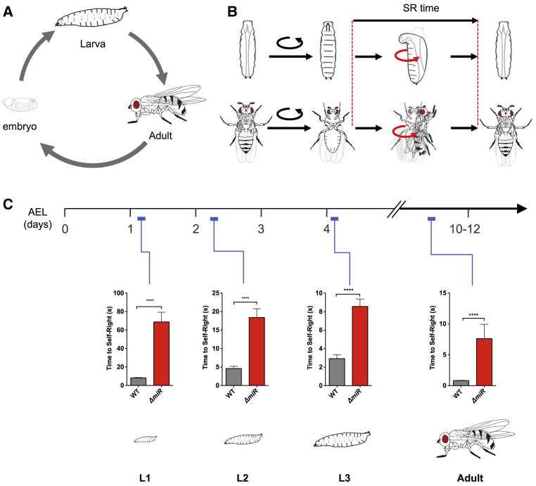
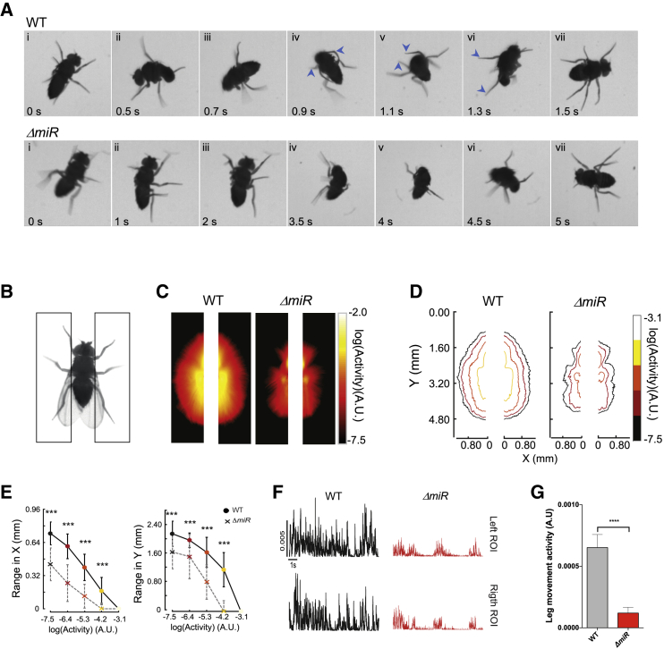
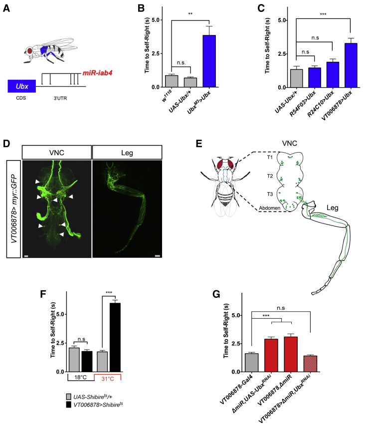
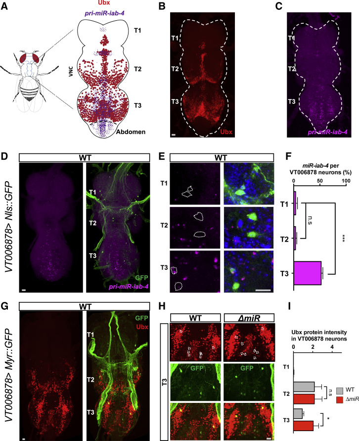
Movement is the main output of the nervous system. It emerges during development to become a highly coordinated physiological process essential to survival and adaptation of the organism to the environment. Similar movements can be observed in morphologically distinct developmental stages of an organism, but it is currently unclear whether or not these movements have a common molecular cellular basis. Here we explore this problem in Drosophila, focusing on the roles played by the microRNA (miRNA) locus miR-iab4/8, which we previously showed to be essential for the normal corrective response displayed by the fruit fly larva when turned upside down (self-righting). Our study shows that miR-iab4 is required for normal self-righting across all three Drosophila larval stages. Unexpectedly, we also discover that this miRNA is essential for normal self-righting behavior in the adult fly, an organism with different morphology, neural constitution, and biomechanics. Through the combination of gene expression, optical imaging, and quantitative behavioral approaches, we provide evidence that miR-iab4 exerts its effects on adult self-righting behavior in part through repression of the Hox gene Ultrabithorax (Ubx) in a specific set of adult motor neurons, the NB2-3/lin15 neurons. Our results show that miRNA controls the function, rather than the morphology, of these neurons and demonstrate that post-developmental changes in Hox gene expression can modulate behavior in the adult. Our work reveals that a common miRNA-Hox genetic module can be re-deployed in different neurons to control functionally equivalent movements in biomechanically distinct organisms and describes a novel post-developmental role of the Hox genes in adult neural function.

Keywords: Drosophila; Hox genes; Ubx; Ultrabithorax; adult/larva; behavior; microRNA; motor neuron; movement; self-righting.


Conflict of interest statement

The authors declare no competing interests.

Figures

Figure 1
Figure 1
Removal of miR-iab4/iab8 Disrupts Larval and Adult Self-Righting Behavior (A) Drosophila melanogaster life cycle. (B) Diagram of SR behavioral response in larvae (top) and adults (bottom). (C) Quantification of the time required for the successful completion of the SR behavior along larval stages and in the adult (mean ± SEM; N = 63–70 larvae for L1, 27–28 for L2, and 25 for L3, and N = 49–54 adult flies) in wild-type controls (w1118, gray) and miR-iab4/iab8 mutants (ΔmiR, red). Analysis of SR behavior throughout development shows that ΔmiR mutants have defects across larval stages and in the adult. A nonparametric Mann-Whitney U test was performed to compare treatments; ∗∗∗p < 0.001. AEL, after egg laying. (NB: Experiments in adult flies were conducted on wingless specimens, but similar results were obtained using different anesthesia methodologies; STAR Methods.) See also Figures S1 and S3 and Videos S1 and S2.
Figure 2
Figure 2
Effects of miRNA Regulation on Leg Movement (A) Description of self-righting (SR) movement in wild-type (top, WT) and miRNA mutant (bottom, ΔmiR) adult flies. Manual video analysis shows that SR behavior in adult WT flies involves several components, including (1) detection of abnormal (upside-down) body orientation, (2) horizontal stirring of legs and body, (3) attempts to grab substrate, (4) coordinated movement of left and right third legs anteriorly until substrate is grabbed, (5) lifting of body by the third legs, (6) tilt of the whole body forward, and (7) return to normal position. (NB: Experiments in adult flies were conducted on wingless specimens; see STAR Methods and Figure 1 legend.) See also Videos S1 and S2. (B–G) Quantification of leg movement levels in WT control (w1118) and ΔmiR flies. Schematic of the paradigm and the regions of interest (black rectangles) drawn to quantify leg movements (STAR Methods) (B). Average heatmaps of leg movements (C) and their corresponding contours (D), and range of movements as defined by the movement contours, in azymuth (right) and elevation (left) axis (E) in ΔmiR flies compared to WT (mean ± SD; two series of experiments, each with N = 10–12 individuals). Color code indicates amplitude of movement, with warm colors representing high levels. Quantification of the leg movements in the ROIs as a function of time (F and G) (mean ± SEM; two independent series of experiments, each with N = 10–12 individuals). Non-parametric Mann-Whitney U test was performed to compare groups; ∗∗∗p < 0.001 and ∗∗∗∗p < 0.001. A.U., arbitrary units. See also Figures S2 and S4.
Figure 3
Figure 3
miRNA-Dependent Ubx Regulation in Ventral VT006878/ventral lin15 Motor Neurons (NB2-3/lin15) Underlies Adult SR Behavior Roles of specific motor neuron subpopulations in SR behavior. (A) The Hox gene Ubx is expressed in the third thoracic region (blue); previous work (see main text) showed that miR-iab4 regulates Ubx expression via specific target sites in Ubx 3′UTR sequences. (B) Quantification of SR behavior in adult flies overexpressing Ubx within its natural expression domain (UbxM3>Ubx: w; UAS-Ubx/+; UbxM3-GAL4/+) shows that upregulation of Ubx is sufficient to cause an adult SR defect (mean ± SEM; n = 19–25). (C) Ubx overexpression in the VT006878/lin15 motor neurons innervating T3 legs phenocopies SR abnormal response (VT006878>Ubx: w; UAS-Ubx/+; VT006878-GAL4/+) (mean ± SEM; three series, each with N = 13–15 flies). (D) Confocal images of VT006878 > Myr::GFP (w; UAS-myr::GFP/+; VT006878-GAL4/+) in the VNC (left) and leg (right); arrows show cell bodies. The projection is displayed to show a maximum of cell bodies by preventing neurite projections. (E) Diagram describing the pattern of VT006878-Gal4 expression in the adult VNC and T3 leg. (F) Blocking neural activity in VT006878 neurons (VT006878>Shibirets: w; +; VT006878-GAL4,+/+,UAS-Shibirets) leads to defects in adult SR response (mean ± SEM; N = 57–64 flies). (G) In ΔmiR adult flies, RNAi-mediated decrease of Ubx expression within the VT006878 domain rescues the SR phenotype (mean ± SEM; N = 41 flies). VT006878, ΔmiR (w; +; VT006878-Gal4,ΔmiR/+,ΔmiR), ΔmiR,UAS-UbxRNAi (w; +; +,ΔmiR/UAS-UbxRNAi,ΔmiR), VT006878>ΔmiR,UbxRNAi (w; +; VT006878-Gal4,ΔmiR/UAS-UbxRNAi,ΔmiR). (NB: Experiments in adult flies were conducted on wingless specimens; see STAR Methods and Figure 1 legend.) Scale bars for anatomic images in (D), 10 μm. Non-parametric Mann-Whitney U (B, C, and F) and one-way ANOVA with the post hoc Tukey-Kramer (G) tests were performed to compare treatments; p > 0.05 (non-significant; n.s.), p < 0.05, and ∗∗∗p < 0.001. See also Figures S5 and S6.
Figure 4
Figure 4
Expression Analysis of Ubx Protein and miR-iab-4 in the Adult VNC (A) Schematic diagram of the adult VNC showing areas of miR-iab-4 (magenta) and Ubx protein (red) expression. (B) Expression of Ubx protein within the VNC shows highest signal in the T3 ganglion, but signal is also detectable in T2 and a much lower level in T1. (C) miR-iab-4 expression in the VNC shows highest level of expression in the T3 ganglion. (D) miR-iab-4 expression profile in VT006878-positive neurons. VT006878 neurons labeled by GFP (VT006878>Nls::GFP: w; UAS-Nls::GFP/+; VT006878-Gal4/+). (E and F) Quantification of miR-iab4 signal (magenta) within the VT006878 domain (green) along different ganglia of the VNC shows a significant increase of miRNA signal in the T3 ganglion (blue, DAPI) (mean ± SEM; n = 7 VNCs). (G) Expression of Ubx protein (red) is detected within the VT006878 domain labeled by GFP (green). VT006878 neurons labeled by GFP (VT006878>Myr::GFP: w; UAS-Myr::GFP/+;VT006878-Gal4/+). (H) Expression pattern of Ubx protein within the VT006878 domain in the T3 region of the VNC in WT (w; UAS-Myr::GFP/+; VT006878-Gal4/+) and miRNA mutants (ΔmiR: w; UAS-Myr::GFP/+; VT006878-Gal4, ΔmiR/+, ΔmiR). A significant increase in Ubx protein expression is observed in the T3 ganglion of mutant adult flies; in contrast, comparison of normal and miRNA mutant flies shows no significant differences in Ubx expression in the T2 ganglion (mean ± SEM; n = 7–13 VNCs). Scale bars for anatomic images, 10 μm. Non-parametric Mann-Whitney U and one-way ANOVA with the post hoc Tukey-Kramer tests were performed to compare treatments; p > 0.05 (not significant; n.s.), p < 0.05, and ∗∗∗p < 0.001. See also Figure S7.
Figure 5
Figure 5
Effects of miRNA Mutation on the Morphology and Function of VT006878/ventral lin15 Motor Neurons (NB2-3/lin15) (A) Image of hind (T3) leg showing VT006878-positive neuronal projections labeled by GFP (VT006878>Myr::GFP). VT006878 neurons innervate the coxa, trochanter, femur, tibia, and tarsus segments. (B) Projections of VT006878 neurons into the coxa, trochanter, femur (left), tibia (middle), and tarsus (right) of wild-type (WT; w; UAS-Myr::GFP/+; VT006878-Gal4/+) and miRNA mutants (ΔmiR; w; UAS-MyrGFP/+; VT006878-Gal4, ΔmiR/+, ΔmiR) specimens show no significant differences across genotypes. For each segment: medial view, left; cross-section, right (mean ± SEM; N = 9 flies per genotype). (C and D) Quantification of VT006878 projections in the segments shows no significant effects of the miRNA system on VT006878/lin15 morphology (dashed line indicates the plane of a cross-section shown at the right of each segment figures; arrowheads highlight motor neuron projections analyzed) (mean ± SEM; N = 8 flies per genotype). (E and F) Varicosity puncta of VT006878 projections in WT and ΔmiR femur. Note the significant reduction in puncta observed in miRNA mutants and the effect caused by Ubx RNAi (ΔmiR,UbxRNAi: w; UAS-myr::GFP/+; VT006878-Gal4, ΔmiR/ UAS-UbxRNAi, ΔmiR) treatment within the VT006878 domain in miRNA mutants, which rescues the normal number of puncta as observed in WT samples (mean ± SEM; N = 10–12 flies per genotype). (G) Schematic representation of the preparation used for calcium activity recordings (top) and the scanned T3 segment (bottom). (H and I) Calcium activity of VT006878 neuron somata and projections within VNC. Representative image for high-resolution morphology (512 × 512) and activity (64 × 64) scans (colored ROIs are detected semi-automatically by Igor software from calcium activity traces in those areas) (left) and an example of calcium activity traces within an ROI (i.e., ROI labeled by a star) reported by GCAMP6m in time indicated by standard normalized fluorescence (SD) (right) (H). Average amplitude, representing area under the curve of the time series, averaged over ROIs (I) of WT (w; UAS-GCAMP6m/+; VT006878-Gal4/+), miRNA mutants (ΔmiR; w; UAS-GCAMP6m/+; VT006878-Gal4, ΔmiR/ +, ΔmiR), and rescue (ΔmiR,UbxRNAi: w; UAS-GCAMP6m/+; VT006878-Gal4, ΔmiR/ UAS-UbxRNAi, ΔmiR) flies (mean ± SEM; N = 5–6 flies per genotype). Non-parametric Mann-Whitney U (C and D) and one-way ANOVA with the post hoc Tukey-Kramer (F and I) tests were performed to compare treatments; p > 0.05 (non-significant; n.s.), p < 0.05, ∗∗p < 0.01, and ∗∗∗p < 0.001. Scale bars for anatomic images, 10 μm.
Figure 6
Figure 6
Conditional Increase of Ubx Expression after Development Is Completed Is Sufficient to Alter Adult Behavior (A) Conditional expression experiment in which Ubx protein is upregulated only once development has been completed. Graphic representation of Gal4 and Gal80 activities over developmental time. NB: At 18°C, Gal80ts represses Gal4 activity; at 31°C, the Gal80ts role is inactivated, allowing for VT006878-Gal4-mediated induction of Ubx (green) in ventral lin15 motor neurons. Maximal induction is achieved approximately 4 days after eclosion (Ti). (B) SR behavior test (STAR Methods) performed at Ti reveals that post-developmental induction of Ubx in VT006878 neurons Tub-Gal80ts; VT006878 > Ubx (w; UAS-Ubx/+; Tub-Gal80ts,+/ +, VT006878-Gal4) (blue) is sufficient to cause SR defects in comparison to control line Tub-Gal80ts;VT006878 > Nls::GFP (w; UAS- Nls::GFP /+; Tub-Gal80ts,+/ +, VT006878-Gal4) (mean ± SEM; N = 19–25 flies). A non-parametric Mann-Whitney U test was performed to compare treatments; ∗∗∗p < 0.001. (C and D) Control treatment for the conditional expression of Ubx in adult Drosophila. Graphic representation of Gal4 and Gal80 activities over developmental time (C). At 18°C, Gal80ts represses Gal4 activity, thus blocking VT006878-Gal4-mediated induction of Ubx in VT006878/ventral lin15 neurons. Under Gal80-mediated repression, there is no induction of Ubx expression in VT006878 cells and no statistically significant changes in SR times are observed when comparing the experimental line Tub-Gal80ts;VT006878; > Ubx (blue) with the control line VT006878;Tub-Gal80ts>Nls::GFP (gray) (mean ± SEM; N = 19–25 flies) (D). A non-parametric Mann-Whitney U test was performed to compare treatments; ∗∗∗p < 0.001. (NB: Experiments in adult flies were conducted on wingless specimens; see STAR Methods and Figure 1 legend.)
Figure 7
Figure 7
Concept Diagram Comparing the Current Understanding of the Neural Basis of Self-Righting Behavior in the Drosophila Larva and Adult (A) Tracings of the larval body wall muscles and lateral transverse muscles 1/2 motor neurons (LT1/2 MNs) [21], projections (green) in the abdominal segments (left), and the illustration of body wall muscles innervated by the motor neurons located in an abdominal hemisegment (A3–A5). In the larva, LT1/2 motor neurons innervate LT1 and LT2 muscles in the body wall; previous work showed that miR-iab4 and Ubx play a particularly important role in the function of these neurons in abdominal segments A3 to A5. (B) In the adult, evidence presented in this study indicates that the miR-iab4::Ubx module is important for normal activity of the VT006878/ ventral lin15 motor neurons in the third thoracic segment. These motor neurons extend complex projections into different leg muscles including the coxa, trochanter, femur, and tibia muscles. The muscles are labeled as described previously [55]: Talm, tarsus levator muscle; tadm, tarsus depressor muscle; tarm, tarsus reductor muscle; tilm, tibia levator muscle; tidm, tibia depressor muscle; tirm, tibia reductor muscle; fedm, femur depressor muscle; ferm, femur reductor muscle; trlm, trochanter levator muscle; trdm, trochanter depressor muscle. The bars to the left of (A) and (B) represent the anatomical size of the three main segments: head (H, red), thorax (T, magenta), and abdomen (A, orange).

Similar articles

Cited by

References

    1. Sherrington C.S. Yale University Press; 1906. The Integrative Action of the Nervous System, Edition.
    1. Sherrington C.S. Some aspects of animal mechanism. Science. 1922;56:345–355. - PubMed
    1. Landmesser L.T., O’Donovan M.J. Activation patterns of embryonic chick hind limb muscles recorded in ovo and in an isolated spinal cord preparation. J. Physiol. 1984;347:189–204. - PMC - PubMed
    1. Saint-Amant L., Drapeau P. Time course of the development of motor behaviors in the zebrafish embryo. J. Neurobiol. 1998;37:622–632. - PubMed
    1. Crisp S., Evers J.F., Fiala A., Bate M. The development of motor coordination in Drosophila embryos. Development. 2008;135:3707–3717. - PMC - PubMed

Publication types

MeSH terms

LinkOut - more resources