

The Effect of Oblique Magnetic Resonance Imaging on Surgical Decision Making for Patients Undergoing an Anterior Cervical Discectomy and Fusion for Cervical Radiculopathy
- PMID: 31328096
- PMCID: PMC6625711
- DOI: 10.14444/6041
The Effect of Oblique Magnetic Resonance Imaging on Surgical Decision Making for Patients Undergoing an Anterior Cervical Discectomy and Fusion for Cervical Radiculopathy
Abstract
Background: The purpose of this study was to determine if oblique magnetic resonance imaging (MRI) sequences affect the surgical treatment recommendations for patients with cervical radiculopathy.
Methods: In this cohort study consecutive clinical cases of persistent cervical radiculopathy requiring surgical intervention were randomized, blinded, and reviewed by 6 surgeons. Initially each surgeon recommended treatment based on the history, physical examination, and axial, coronal and sagittal preoperative magnetic resonance (MR) images; when reviewing the cases the second time, the surgeons were provided oblique MR images. This entire process was then repeated after 2 months. Change in surgical recommendation, interobserver and intraobserver reliability and the average number of levels fused was determined.
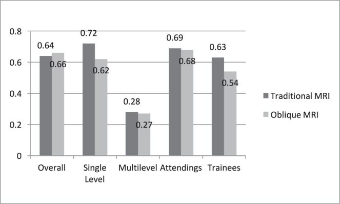
Results: The addition of the oblique images resulted in the surgical recommendation being altered in 49.2% (59/120) of cases; however, the addition of oblique images did not substantially improve the interobserver reliability of the treatment recommendation (κ = .57 versus.57). Similarly, the overall intraobserver reliability using only traditional MRI sequences (κ = .64) was only slightly improved by the addition of oblique images (κ = .66). Lastly, the addition of oblique images did not change the average number of levels fused (traditional MRI = 1.38, oblique MRI = 1.41, P = .53), or the total number of 3-level fusions recommended (6 versus 6, P = 1.00).
Conclusions: The additional oblique images resulted in a change to the surgical plan in almost 50% of cases; however, it had no substantial effect on the reliability of surgical decision making. Further studies are needed to see if this alteration in treatment affects clinical outcomes.
Level of evidence: 3.
Keywords: ACDF; cervical, radiculopathy; fusion; magnetic resonance imaging.
Conflict of interest statement
Disclosures and COI: No funds were received in support of this work. The authors disclose no direct or indirect conflicts of interest in the preparation of this manuscript. No pharmaceutical or medical devices were utilized during this study. No benefits in any form have been or will be received from a commercial party related directly or indirectly to the subject of this manuscript. This included the National Institutes of Health (NIH); Wellcome Trust; Howard Hughes Medical Institute (HHMI). The authors had password-protected access to the data with nonessential patient demographics deidentified. Ethics Board Review Statement: This study was approved by the Institutional Review Board at Thomas Jefferson University Hospital. Each author certifies that our institution approved the human protocol for this investigation and that all investigations were conducted in conformity with ethical principles of research.
Figures


References
-
- Radhakrishnan K, Litchy WJ, O'Fallon WM, Kurland LT. Epidemiology of cervical radiculopathy. A population-based study from Rochester, Minnesota, 1976 through 1990. Brain. 1994;117(Pt 2):325–335. - PubMed
-
- Henderson CM, Hennessy RG, Shuey HM, Jr, Shackelford EG. Posterior-lateral foraminotomy as an exclusive operative technique for cervical radiculopathy: a review of 846 consecutively operated cases. Neurosurgery. 1983;13(5):504–512. - PubMed
-
- Rhee JM, Yoon T, Riew KD. Cervical radiculopathy. J Am Acad Orthop Surg. 2007;15(8):486–494. - PubMed
-
- Modic MT, Masaryk TJ, Ross JS, Mulopulos GP, Bundschuh CV, Bohlman H. Cervical radiculopathy: value of oblique MR imaging. Radiology. 1987;163(1):227–231. - PubMed
LinkOut - more resources
Full Text Sources