. 2019 Oct;23(10):6708-6719.
doi: 10.1111/jcmm.14549. Epub 2019 Jul 21.

LncRNA MEG3 inhibits cell proliferation and induces apoptosis in laryngeal cancer via miR-23a/APAF-1 axis

Affiliations

LncRNA MEG3 inhibits cell proliferation and induces apoptosis in laryngeal cancer via miR-23a/APAF-1 axis

Xiaowen Zhang et al. J Cell Mol Med. 2019 Oct.

Abstract

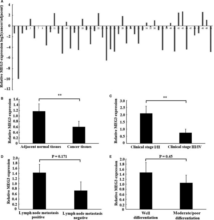
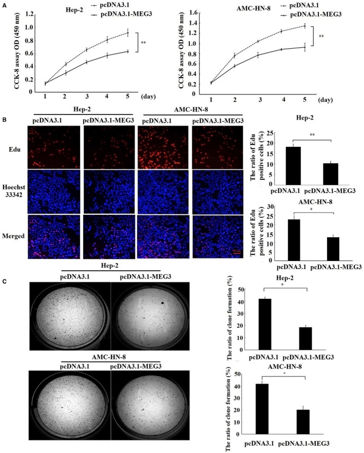
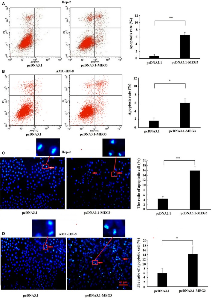
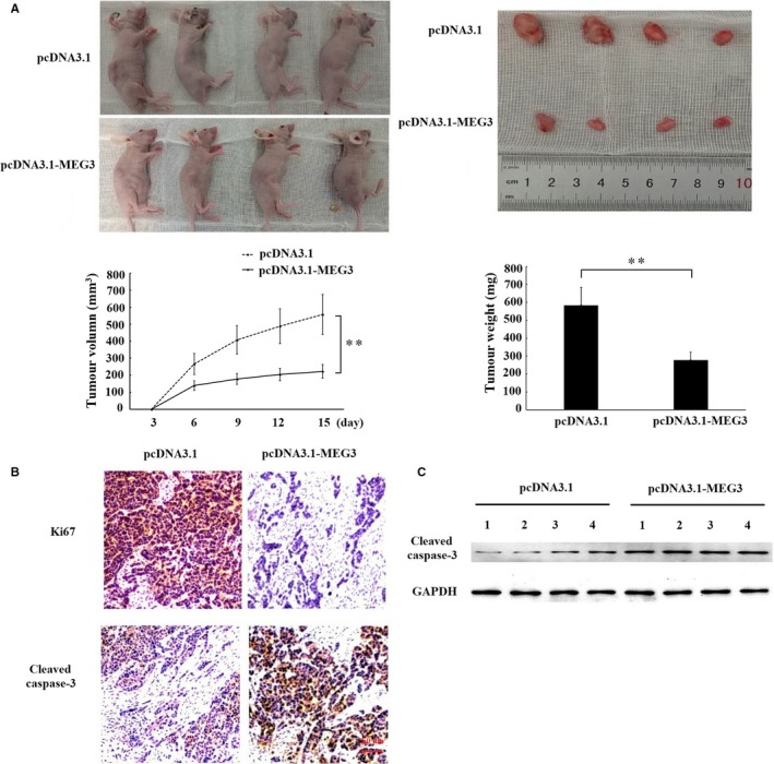
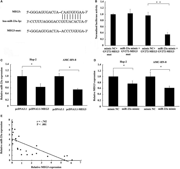
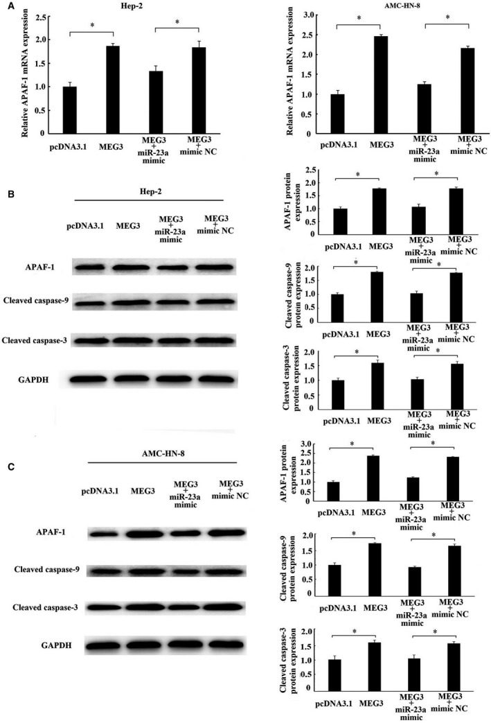
Long non-coding RNA (LncRNA) MEG3 serves a regulatory role in the progression of several types of cancer, but the role of MEG3 in laryngeal cancer is still unknown. The aim of this study was to explore the regulatory role and mechanism of MEG3 in laryngeal cancer. MEG3 expression in 50 laryngeal cancer tissue samples was detected by reverse transcription-quantitative polymerase chain reaction (RT-qPCR). The effects of MEG3 overexpression on laryngeal cancer cells were investigated in vitro and in vivo. The mechanism of competitive endogenous RNA (ceRNA) was validated through luciferase reporter assay, RT-qPCR and Western blotting. MEG3 was down-regulated in laryngeal cancer tissues, and the low MEG3 expression was associated with advanced clinical stage. Additionally, MEG3 overexpression inhibited the proliferation and induced the apoptosis of laryngeal cancer cells in vitro and in vivo. Particularly, MEG3 bound to miR-23a specifically and a reciprocal negative regulation existed between miR-23a and MEG3. Moreover, MEG3 up-regulated apoptotic protease activating factor-1 (APAF-1), a known miR-23a's target, thereby leading to the activation of caspase-9 and caspase-3. Meanwhile, these activated effects were rescued by miR-23a overexpression. In conclusion, the present study demonstrated that MEG3 functions as a novel tumour suppressive LncRNA in laryngeal cancer for the first time. Furthermore, MEG3 may act as a ceRNA to regulate APAF-1 expression by competitive binding to miR-23a, thereby regulating the progression of laryngeal cancer.

Keywords: apoptotic protease activating factor-1; competitive endogenous RNA; laryngeal cancer; maternally expressed gene 3; miRNA-23a.


Conflict of interest statement

The authors have no conflict of interest.

Figures

Figure 1
Figure 1
Expression of MEG3 in laryngeal cancer tissues. The relative expression of MEG3 was examined using RT‐qPCR in 50 patients with laryngeal cancer. A, The relative MEG3 expression was detected in each of laryngeal cancer tissues and matched normal tissues. Data were presented as fold change of laryngeal cancer tissues relative to adjacent normal regions (B) Relative MEG3 expression level in laryngeal cancer tissues and adjacent normal regions. (C), (D) and (E) the statistical analysis of the association between MEG3 expression and clinical stages, lymph node metastasis and differentiation. Data were presented as mean ± SEM, **P < .01
Figure 2
Figure 2
Effects of MEG3 on the proliferation of laryngeal cancer cells in vitro. Hep‐2 and AMC‐HN‐8 cells were transfected with MEG3 plasmid (pcDNA3.1‐MEG3) and empty vectors (pcDNA3.1). A, CCK‐8 assay was performed to measure the cell proliferation for 5 consecutive days. B, EdU assay was performed to evaluate the cell proliferation. Magnification: ×200, scale bars: 20 μm. C, Clone formation assay was performed to detect the ability of clone formation in Hep‐2 and AMC‐HN‐8 cells. The data were obtained from three independent experiments and presented as mean ± SEM, *P < .05.**P < .01
Figure 3
Figure 3
Effects of MEG3 on the apoptosis of laryngeal cancer cells. Hep‐2 and AMC‐HN‐8 cells were transfected with MEG3 plasmid (pcDNA3.1‐MEG3) and empty vectors (pcDNA3.1). Flow cytometry was employed to detect the apoptotic rates of cells in Hep‐2 (A) and AMC‐HN‐8 cells (B), cells in the lower right quadrant represent apoptosis cells. Hoechst staining assay was performed to assess cell apoptosis in Hep‐2 (C) and AMC‐HN‐8 cells (D). Hoechst‐positive nuclei (apoptotic nuclei) showed dense and high‐density fluorescence, as indicated by arrows, and the percentage of Hoechst‐positive nuclei per optical field (at least 10 fields) was counted. Magnification: ×200, scale bars: 20 μm. The data were obtained from three independent experiments and presented as mean ± SEM, *P < .05, **P < .01
Figure 4
Figure 4
The effects of MEG3 on the growth of laryngeal cancer cells in vivo. Hep‐2 cells transfected with empty vector or MEG3 plasmid were injected into nude mice (n = 4/group). A, Tumour volumes were measured every 3 d after injection. At 15 d after injection, xenograft tumours were harvested and weighed. B, Immunohistochemistry assay was performed to assess the protein levels of Ki67 and cleaved caspase‐3. Magnification: ×400, scale bars: 50 μm. C, Western blotting was employed to measure protein level of cleaved caspase‐3 in xenograft tumour tissues. The data were obtained from three independent experiments and presented as mean ± SEM, ** P < .01
Figure 5
Figure 5
The interaction of MEG3 with miR‐23a. A, The binding site of miR‐23a on MEG3 transcript. B, Luciferase reporter gene assay validates the interaction between MEG3 and miR‐23a in Hep‐2 cells. GV272‐MEG3, luciferase vector of MEG3; GV272‐MEG3‐mut, MEG3 mutant vector. C, The effect of MEG3 overexpression on miR‐23a expression in Hep‐2 and AMC‐HN‐8 cells. D, The effect of miR‐23a overexpression on MEG3 expression in Hep‐2 and AMC‐HN‐8 cells. E, A negative relationship between MEG3 and miR‐23a expression existed in laryngeal cancer tissues. The data were presented as mean ± SEM, *P < .05, **P < .01
Figure 6
Figure 6
Regulation of APAF‐1 by MEG3 was mediated by miR‐23a. MEG3 plasmid (pcDNA3.1‐MEG3) and empty vector (pcDNA3.1) alone or together with miR‐23a mimics or miR‐23a‐negative control (mimic NC) were transfected into Hep‐2 and AMC‐HN‐8 cells. A, The mRNA expression of APAF‐1 in Hep‐2 and AMC‐HN‐8 cells was detected by RT‐qPCR. Western blotting analysed the protein levels of APAF‐1, cleaved caspase‐9 and cleaved caspase‐3 in Hep‐2 (B) and AMC‐HN‐8 cells (C). The data were obtained from three independent experiments and presented as mean ± SEM. * P < .05

References

    1. Siegel RL, Miller KD, Jemal A. Cancer statistics, 2016. CA Cancer J Clin. 2016;66:7‐30. - PubMed
    1. Menach P, Oburra HO, Patel A. Cigarette smoking and alcohol ingestion as risk factors for laryngeal squamous cell carcinoma at Kenyatta National Hospital, Kenya. Clin Med Insights Ear Nose Throat. 2012;5:17‐24. - PMC - PubMed
    1. Garavello W, Turati F, Bosetti C, et al. Family history of cancer and the risk of laryngeal cancer: a case‐control study from Italy and Switzerland. Int J Cancer. 2012;130:665‐670. - PubMed
    1. Todorova TA, Jordanov SH, Stancheva GS, et al. Mutational status of CDKN2A and TP53 genes in laryngeal squamous cell carcinoma. Pathol Oncol Res. 2015;21:413‐421. - PubMed
    1. Fanjul‐Fernández M, Quesada V, Cabanillas R, et al. Cell‐cell adhesion genes CTNNA2 and CTNNA3 are tumour suppressors frequently mutated in laryngeal carcinomas. Nat Commun. 2013;4:2531. - PubMed

Publication types

MeSH terms