. 2019 Oct;23(10):6785-6796.
doi: 10.1111/jcmm.14559. Epub 2019 Jul 21.

Glucocorticoid receptor inhibits Müller glial galectin-1 expression via DUSP1-dependent and -independent deactivation of AP-1 signalling

Affiliations

Glucocorticoid receptor inhibits Müller glial galectin-1 expression via DUSP1-dependent and -independent deactivation of AP-1 signalling

Ikuyo Hirose et al. J Cell Mol Med. 2019 Oct.

Abstract

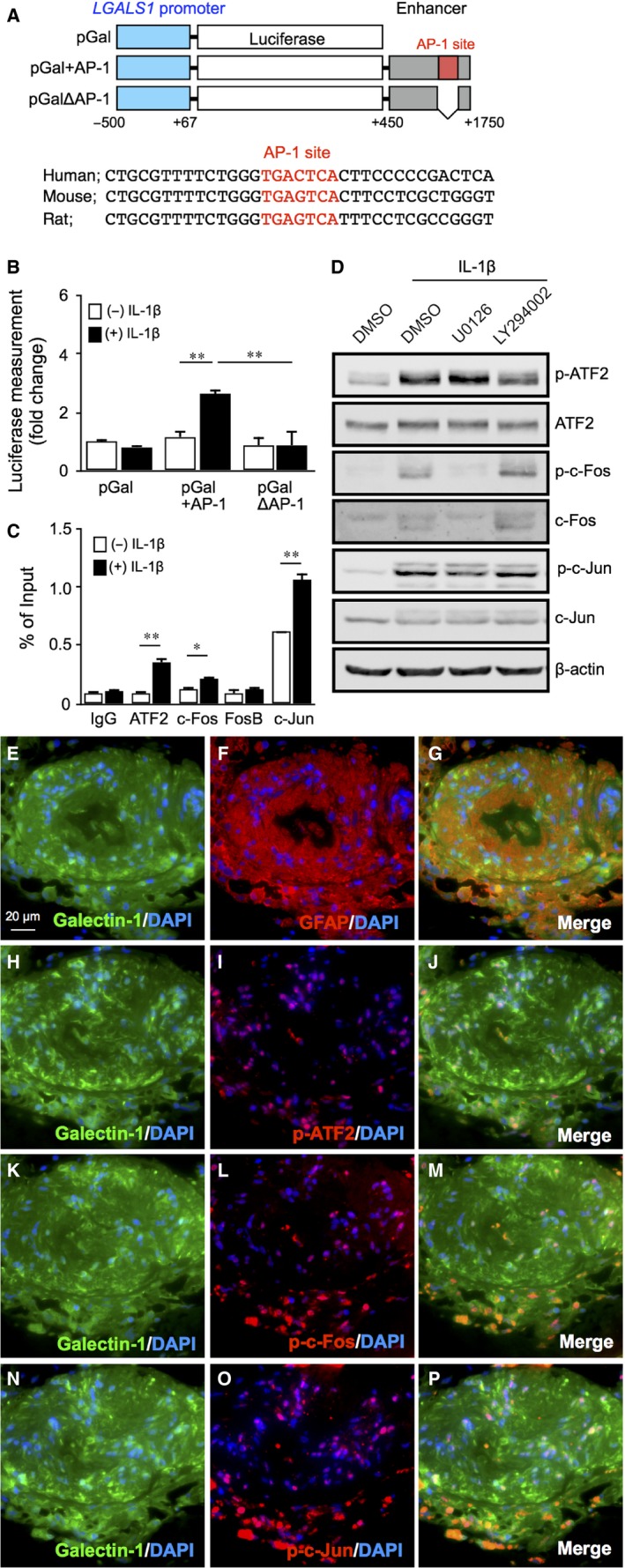
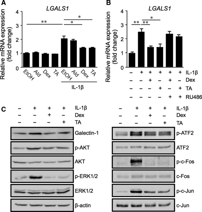
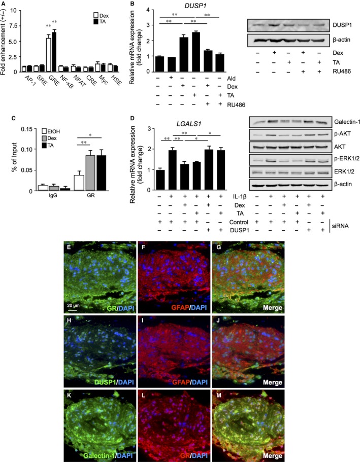
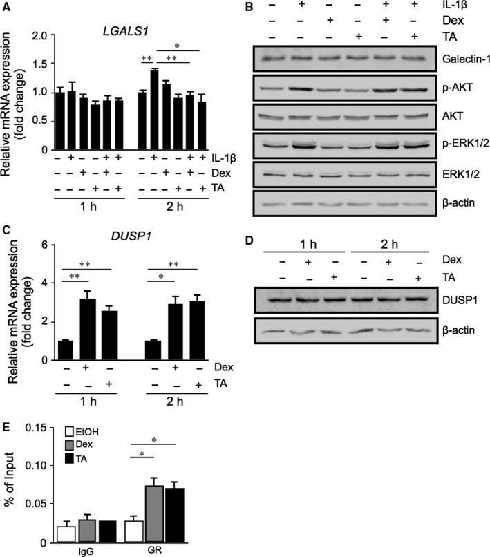
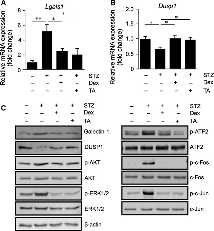
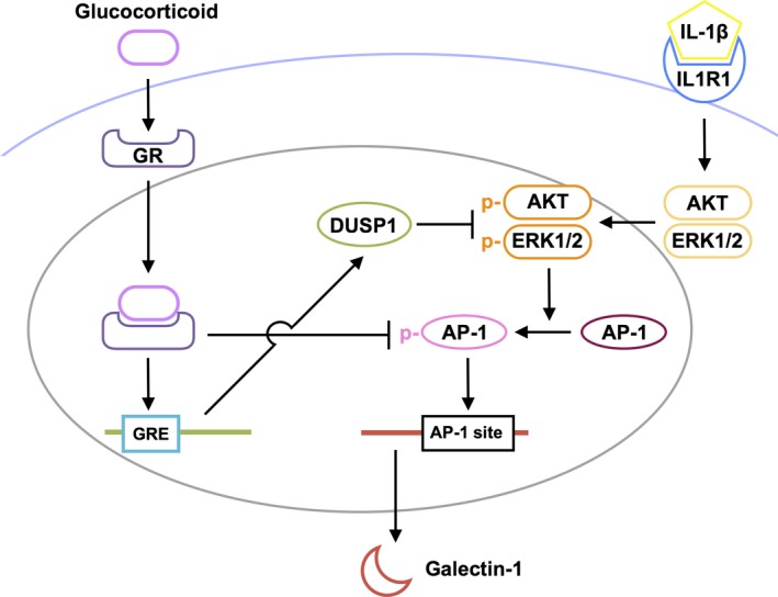
Galectin-1/LGALS1 is a hypoxia-induced angiogenic factor associated with diabetic retinopathy (DR). Recently, we elucidated a hypoxia-independent pathway to produce galectin-1 in Müller glial cells stimulated by interleukin (IL)-1β. Here we revealed glucocorticoid receptor (GR)-mediated inhibitory mechanisms for Müller glial galectin-1/LGALS1 expression. Activator protein (AP)-1 site in the LGALS1 enhancer region, to which activating transcription factor2, c-Fos and c-Jun bind, was shown to be essential for IL-1β-induced galectin-1/LGALS1 expression in Müller cells. Ligand (dexamethasone or triamcinolone acetonide)-activated GR induced dual specificity phosphatase (DUSP)1 expression via the glucocorticoid response element and attenuated IL-1β-induced galectin-1/LGALS1 expression by reducing phosphorylation of these AP-1 subunits following AKT and extracellular signal-regulated kinase (ERK)1/2 deactivation. Moreover, activated GR also caused DUSP1-independent down-regulation of IL-1β-induced LGALS1 expression via its binding to AP-1. Administration of glucocorticoids to mice attenuated diabetes-induced retinal galectin-1/Lgals1 expression together with AKT/AP-1 and ERK/AP-1 pathways. Supporting these in vitro and in vivo findings, immunofluorescence analyses showed co-localization of galectin-1 with GR and phosphorylated AP-1 in DUSP1-positive glial cells in fibrovascular tissues from patients with DR. Our present data demonstrated the inhibitory effects of glucocorticoids on glial galectin-1 expression via DUSP1-dependent and -independent deactivation of AP-1 signalling (transactivation and transrepression), highlighting therapeutic implications for DR.

Keywords: Müller glia; activator protein-1; diabetic retinopathy; dual specificity phosphatase 1; galectin-1; glucocorticoid receptor; interleukin-1β; transactivation; transrepression.


Conflict of interest statement

The authors declare no competing financial interests.

Figures

Figure 1
Figure 1
Requirement of AKT‐ and ERK1/2‐dependent AP‐1 activity in IL‐1β‐induced LGALS1 expression in Müller glial cells. A, Upper, schematic representation of LGALS1 promoter‐ and enhancer‐driven luciferase constructs. Lower, sequence comparison between human, mouse and rat LGALS1 enhancer regions spanning AP‐1 site (red). B, AP‐1 site in the LGALS1 enhancer region was required for IL‐1β‐enhanced LGALS1 promoter‐driven luciferase activity. Transfected Müller glial cells were stimulated with IL‐1β (10 ng/mL) for 24 h and assayed for luciferase activity. C, Müller glial cells were stimulated with IL‐1β (10 ng/mL) for 1 h before harvest of samples. Binding of ATF2, c‐Fos, FosB and c‐Jun to AP‐1 site in the LGALS1 enhancer region was analysed by ChIP‐qPCR. *P < .05, **P < .01, n = 4‐6 per group. D, Müller glial cells were pre‐treated with each inhibitor at 10 μmol/L for 30 min before stimulation with IL‐1β (10 ng/mL) for 1 h, and protein levels of phosphorylated and total forms of AP‐1 subunits were analysed. (E‐P) Double labelling of galectin‐1 (green), GFAP (red) and DAPI (blue) (E‐G); galectin‐1 (green), phosphorylated ATF2 (red) and DAPI (blue) (H‐J); galectin‐1 (green), phosphorylated c‐Fos (red) and DAPI (blue) (K‐M); galectin‐1 (green), phosphorylated c‐Jun (red) and DAPI (blue) (N‐P) in fibrovascular tissues excised from human eyes with proliferative DR. Scale bar = 20 μm
Figure 2
Figure 2
Glucocorticoid‐mediated suppression of IL‐1β‐induced galectin‐1/LGALS1 expression with AKT/AP‐1 and ERK/AP‐1 activation in Müller glial cells. A, Müller glial cells were pre‐treated with aldosterone (Ald, 1 μmol/L), dexamethasone (Dex, 1 μmol/L) or triamcinolone acetonide (TA, 1 μmol/L) for 30 min before stimulation with IL‐1β (10 ng/mL) for 24 h, and LGALS1 gene expression levels were analysed. B, Müller glial cells were pre‐treated with the GR antagonist (RU486, 1 μmol/L) for 30 min before application with Dex (1 μmol/L), TA (1 μmol/L) and IL‐1β (10 ng/mL) for 24 h, and LGALS1 gene expression levels were analysed. *P < .05, **P < .01, n = 8 per group. C, Müller glial cells were pre‐treated with Dex or TA at 1 μmol/L for 30 min before stimulation with IL‐1β (10 ng/mL) for 24 h, and protein levels of galectin‐1, phosphorylated and total forms of AKT, ERK1/2 and AP‐1 subunits were analysed
Figure 3
Figure 3
Glucocorticoid‐transactivated DUSP1 expression as an exclusive suppressor of IL‐1β‐induced galectin‐1/LGALS1 expression with AKT and ERK1/2 activation in Müller glial cells. A, Transcriptional factors activated by glucocorticoids. Müller glial cells were transfected with plasmids containing the following response elements or binding sites (AP‐1; SRE, serum response element; GRE; NF‐κB; NFAT, nuclear factor of activated T cells; CRE, cAMP response element; Myc, E‐box DNA‐binding element; HSE, heat shock response element). After 24 h, cells were stimulated with dexamethasone (Dex, 1 μmol/L) or triamcinolone acetonide (TA, 1 μmol/L) for 48 h and assayed for alkaline phosphatase activity. Fold change is relative to vehicle‐treated controls. B, Müller glial cells were pre‐treated with the GR antagonist RU486 (1 μmol/L) for 30 min before application with aldosterone (Ald, 1 μmol/L), Dex (1 μmol/L) or TA (1 μmol/L) for 24 h, and DUSP1 gene and protein expression levels were analysed. n = 6‐8 per group. C, Müller glial cells were treated with Dex (1 μmol/L) or TA (1 μmol/L) for 1 h, and GR binding to GRE in the DUSP1 promoter region was analysed by ChIP‐qPCR. n = 4‐6 per group. D, After DUSP1 knockdown, cells were applied with Dex (1 μmol/L) or TA (1 μmol/L) and 10 ng/mL IL‐1β for 24 h, LGALS1 gene and protein expression levels were analysed. Immunoblots were probed with antibodies against galectin‐1 and phosphorylated and total forms of AKT and ERK1/2. *P < .05, **P < .01, n = 6‐8 per group. (E‐M) Double labelling of GR (green), GFAP (red) and DAPI (blue) (E‐G); DUSP1 (green), GFAP (red) and DAPI (blue) (H‐J); galectin‐1 (green), GR (red) and DAPI (blue) (K‐M) in fibrovascular tissues excised from human eyes with proliferative DR. Scale bar = 20 μm
Figure 4
Figure 4
DUSP1‐independent down‐regulation of LGALS1 expression because of deactivation of AP‐1 signalling via GR binding to AP‐1 in Müller glial cells. A, Müller glial cells were applied with dexamethasone (Dex, 1 μmol/L) or triamcinolone acetonide (TA, 1 μmol/L) and 10 ng/mL IL‐1β for 1 or 2 h, and LGALS1 gene expression levels were analysed. B, Müller glial cells were applied with Dex (1 μmol/L) or TA (1 μmol/L) and 10 ng/mL IL‐1β for 2 h, and protein levels of galectin‐1 and phosphorylated and total forms of AKT and ERK1/2 were analysed. (C,D) Müller glial cells were treated with Dex (1 μmol/L) or TA (1 μmol/L) for 1 or 2 h, and DUSP1 mRNA (C) and protein (D) expression levels were analysed. E, Müller glial cells were applied with Dex (1 μmol/L) or TA (1 μmol/L) and 10 ng/mL IL‐1β for 1 h, and binding of activated GR to the AP‐1/AP‐1 site complex was analysed by ChIP‐qPCR. *P < .05, **P < .01, n = 4‐6 per group
Figure 5
Figure 5
Glucocorticoid‐mediated suppression of diabetes‐induced galectin‐1/Lgals1 expression with AKT/AP‐1 and ERK/AP‐1 activation in the mouse retina. (A‐C) Retinal Lgals1 (A) and Dusp1 (B) expression in mice with STZ‐induced diabetes at 2 mo. Dexamethasone (Dex, 50 pmol/eye) or triamcinolone acetonide (TA, 50 pmol/eye) were injected intravitreally to STZ mice, followed by mRNA (A,B) and protein (C) expression analyses after 24 h. *P < .05, **P < .01, n = 6‐8 per group. C, Immunoblot analyses for galectin‐1, DUSP1 and phosphorylated and total forms of AKT, ERK1/2 and AP‐1 subunits in the retina of diabetic mice treated with Dex or TA
Figure 6
Figure 6
A schema showing the inhibitory effects of ligand‐bound GR on IL‐1β‐induced galectin‐1 expression with AP‐1 signalling in Müller glial cells. Ligand‐bound GR inhibits Müller glial galectin‐1 expression via DUSP1‐dependent (transactivation) and ‐independent (transrepression) deactivation of AKT/AP‐1 and ERK/AP‐1 signal transduction induced by IL‐1β

References

    1. Camby I, Le Mercier M, Lefranc F, Kiss R. Galectin‐1: a small protein with major functions. Glycobiology. 2006;16:137R‐157R. - PubMed
    1. Croci D, Cerliani J, Dalotto‐Moreno T, et al. Glycosylation‐dependent lectin‐receptor interactions preserve angiogenesis in anti‐VEGF refractory tumors. Cell. 2014;156:744‐758. - PubMed
    1. Yang N, Zhang W, He T, Xing Y. Silencing of galectin‐1 inhibits retinal neovascularization and ameliorates retinal hypoxia in a murine model of oxygen‐induced ischemic retinopathy. Exp Eye Res. 2017;159:1‐15. - PubMed
    1. Kanda A, Noda K, Saito W, Ishida S. Aflibercept traps galectin‐1, an angiogenic factor associated with diabetic retinopathy. Sci Rep. 2015;5:17946. - PMC - PubMed
    1. Kanda A, Dong Y, Noda K, Saito W, Ishida S. Advanced glycation endproducts link inflammatory cues to upregulation of galectin‐1 in diabetic retinopathy. Sci Rep. 2017;7:16168. - PMC - PubMed

Publication types

MeSH terms