. 2019 Jul 22;14(7):e0218895.
doi: 10.1371/journal.pone.0218895. eCollection 2019.

Fatty acid and retinol-binding protein: A novel antigen for immunodiagnosis of human strongyloidiasis

Affiliations

Fatty acid and retinol-binding protein: A novel antigen for immunodiagnosis of human strongyloidiasis

Leila Masoori et al. PLoS One. .

Abstract

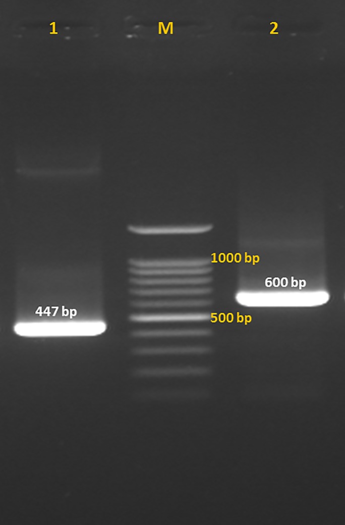
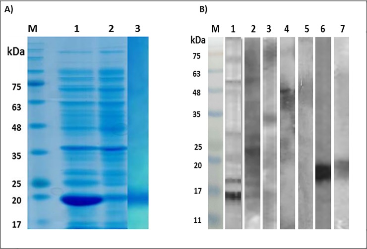
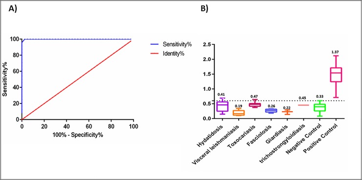
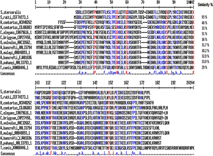
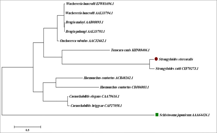
The tenacious human parasitic helminth Strongyloides stercoralis is a significant health problem worldwide. The current lack of a definitive diagnostic laboratory test to rule out this infection necessitates designing more specific diagnostic methods. Fatty acid and retinol-binding protein (FAR) plays a crucial role in the development and reproduction of nematodes. We generated a recombinant form of this protein and determined its applicability for immunodiagnosis of S. stercoralis. The L3 form of S. stercoralis was harvested and used for RNA extraction and cDNA synthesis. The coding sequence of S. stercoralis FAR (SsFAR) was cloned into pET28a(+) vector, expressed in E. coli BL21 and purified. ELISA and immunoblotting were employed to determine the specificity and sensitivity of rSsFAR using a set of defined sera. In addition, we analyzed the phylogenetic relationship of SsFAR with different FAR sequences from other nematodes. The cloned SsFAR had an open reading frame of 447 bp encoding 147 amino acids, with a deduced molecular mass of 19 kD. The SsFAR amino acid sequence was 93% identical to FAR of S. ratti. For differential immunodiagnosis of strongyloidiasis, rSsFAR exhibited 100% sensitivity and 97% specificity. However, cross-reactivity with FAR proteins of other parasites, namely Toxocara canis and Echinococcus granulosus, was noted. Our results provide a novel approach for immunodiagnosis of S. stercoralis infections using rSsFAR with reliable sensitivity and specificity.


Conflict of interest statement

The authors have declared that no competing interests exist.

Figures

Fig 1
Fig 1. Fatty acid and retinol-binding protein (FAR) gene fragments of Strongyloides stercoralis.
Fatty acid and retinol binding protein (FAR) sequence of infective larvae of Strongyloides stercoralis was amplified by conventional PCR and following insertion in pTG19-T/A vector was rechecked. Lane 1: PCR product of the amplification of FAR gene from the cDNA of infective larvae of S. stercoralis; Lane 2: PCR amplification of FAR gene which was inserted into pTG19-T/A vector with M13-specific primers: Lane M: 100 bp DNA ladder (Cinnagen, Tehran, Iran).
Fig 2
Fig 2. SDS-PAGE and immunoblotting of the crude extract, as well as recombinant fatty acid and retinol-binding protein of Strongyloides stercoralis.
Fatty acid and retinol-binding protein (FAR) was expressed in a prokaryotic host and the immunoreactivity of the purified protein was analyzed by immunoblotting. A: Strongyloides stercoralis FAR gene was cloned in pET28a(+) plasmid, transformed in E. coli BL21 and the expression of the rFAR protein was induced by 1mM IPTG within four hours of incubation at 37°C. The bacterial cells were lysed and following centrifugation the contents of the precipitated and the supernatant fractions was analyzed by 15% SDS-PAGE gels for evaluation of the expression of the target protein. The slabs were stained with Coomassie brilliant blue G250. Lane M: Protein molecular weight marker. Lane 1: Supernatant of the lysed bacterial cells showing overexpression of a 19 kDa recombinant protein; Lane 2: Precipitate of the lysed bacterial cells, Lane 3: Purified rFAR. B: The crude extract from infective larvae of S. stercoralis and purified rFAR was immunoblotted with pooled sera from hyper-infected strongyloidiasis patients, as well as toxoariasis and hydatidosis patients. Furthermore, anti-his-tagged antibody was used for characterization of the recombinant protein. M: Protein molecular weight marker, Lane 1–2: Immunoblotting of the crude extract with pooled sera of five strongyloidiasis patients; 3: Immunoblotting of the crude extract with polled sera of two hyper-infected patients; Lane 4: Immunoblotting of the crude extract with toxocariasis patients’; Lane 5: Immunoblotting of the crude extract with pooled sera of hydatidosis patients; Lane 6: Immunoblotting of rFAR protein with anti-his-tagged antibody, Lane 7: Immunoblotting of rFAR protein with pooled sera of strongyloidiasis patients.
Fig 3
Fig 3. Receiver-operating characteristic (ROC) curves and the Cross-reactivity of rFAR sera from patients with other parasitic diseases.
A: The ROC curve was plotted using optical density (OD) obtained in ELISA for 33 serum samples from S. stercoralis-infected patients and 40 healthy controls. The area under the curve (AUC) for recombinant Fatty acid and retinol-binding protein (rFAR) was 0.99. B: Cross-reactivity of rFAR with antibodies against other parasitic diseases was determined by indirect ELISA. Sera from some other parasitic diseases were examined with rFAR-coated ELISA plates and did not show considerable cross-reactivity.
Fig 4
Fig 4. Multiple sequence alignment of fatty acid and retinol-binding protein (FAR) of S. stercoralis.
Fatty acid and retinol-binding protein (FAR) of S. stercoralis and other nematode including S. ratti (CEF70273.1), H. contortus (ACR48262.1, CDJ86883), C. elegans (CAA79616.1), C. briggsae (CAP27058.1), O. volvulus (AAC32662.1), W. bancrofti (AAL33794.1, EJW81696.1), B. malayi (AAB08893.1), B. pahangi (AAL33793.1), T. canis (KHN88406.1) were analyzed by multiple sequence alignment using NCBI protein database, online ClustalW (http://www.clustal.org/clustal2/) and Multalin (http://multalin.toulouse.inra.fr/multalin/) software.
Fig 5
Fig 5. Phylogenetic tree of fatty acid and retinol-binding protein (FAR) of S. stercoralis with other nematodes.
The phylogenetic tree was constructed using amino acids sequences of S. ratti (CEF70273.1), H. contortus (ACR48262.1, CDJ86883), C. elegans (CAA79616.1), C. briggsae (CAP27058.1), O. volvulus (AAC32662.1), W. bancrofti (AAL33794.1, EJW81696.1), B. malayi (AAB08893.1), B. pahangi (AAL33793.1), T. canis (KHN88406.1) obtained from NCBI protein database. The Neighbor-joining phylogenetic tree rooted against the FAR protein sequences of the trematode Schistosoma japonicum was generated using MEGA 5 software (Arizona State University, Tempe, USA).

References

    1. Krolewiecki AJ, Lammie P, Jacobson J, Gabrielli A-F, Levecke B, Socias E, et al. A public health response against Strongyloides stercoralis: time to look at soil-transmitted helminthiasis in full. PLoS Negl Trop Dis. 2013;7(5):e2165 10.1371/journal.pntd.0002165 - DOI - PMC - PubMed
    1. Schär F, Trostdorf U, Giardina F, Khieu V, Muth S, Marti H, et al. Strongyloides stercoralis: global distribution and risk factors. PLoS Negl Trop Dis. 2013;7(7):e2288 10.1371/journal.pntd.0002288 - DOI - PMC - PubMed
    1. Bisoffi Z, Buonfrate D, Montresor A, Requena-Méndez A, Muñoz J, Krolewiecki AJ, et al. Strongyloides stercoralis: a plea for action. PLoS Negl Trop Dis. 2013;7(5):e2214 10.1371/journal.pntd.0002214 - DOI - PMC - PubMed
    1. Nasiri V, Esmailnia K, Karim G, Nasir M, Akhavan O. Intestinal parasitic infections among inhabitants of Karaj City, Tehran province, Iran in 2006–2008. The Korean journal of parasitology. 2009;47(3):265 10.3347/kjp.2009.47.3.265 - DOI - PMC - PubMed
    1. Kia E, Mahmoudi M, Zahabiun F, Meamar A. An evaluation on the efficacy of agar plate culture for detection of Strongyloides stercoralis. Iran J Parasitol. 2007;2(1):29–34.

Publication types

MeSH terms