. 2019 Sep 19;47(16):8581-8594.
doi: 10.1093/nar/gkz606.

Are the intrinsically disordered linkers involved in SSB binding to accessory proteins?

Affiliations

Are the intrinsically disordered linkers involved in SSB binding to accessory proteins?

Min Kyung Shinn et al. Nucleic Acids Res. .

Abstract

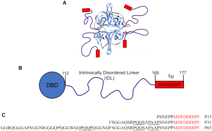
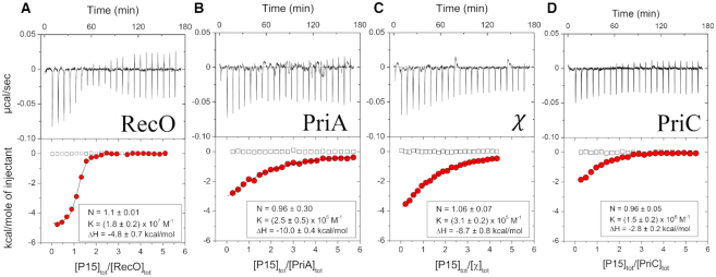
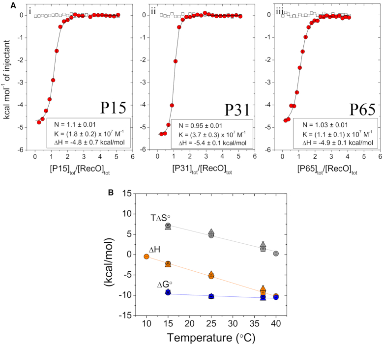
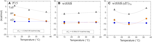
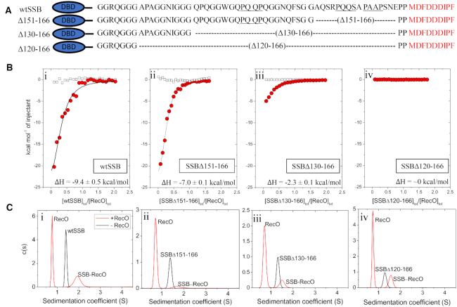
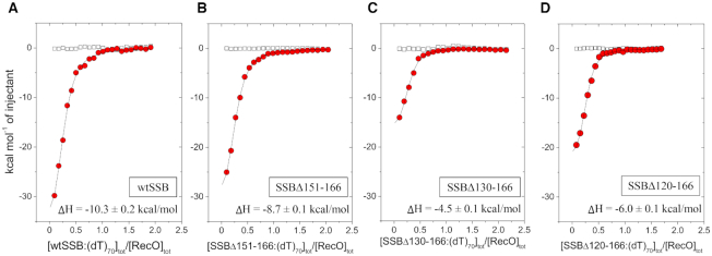
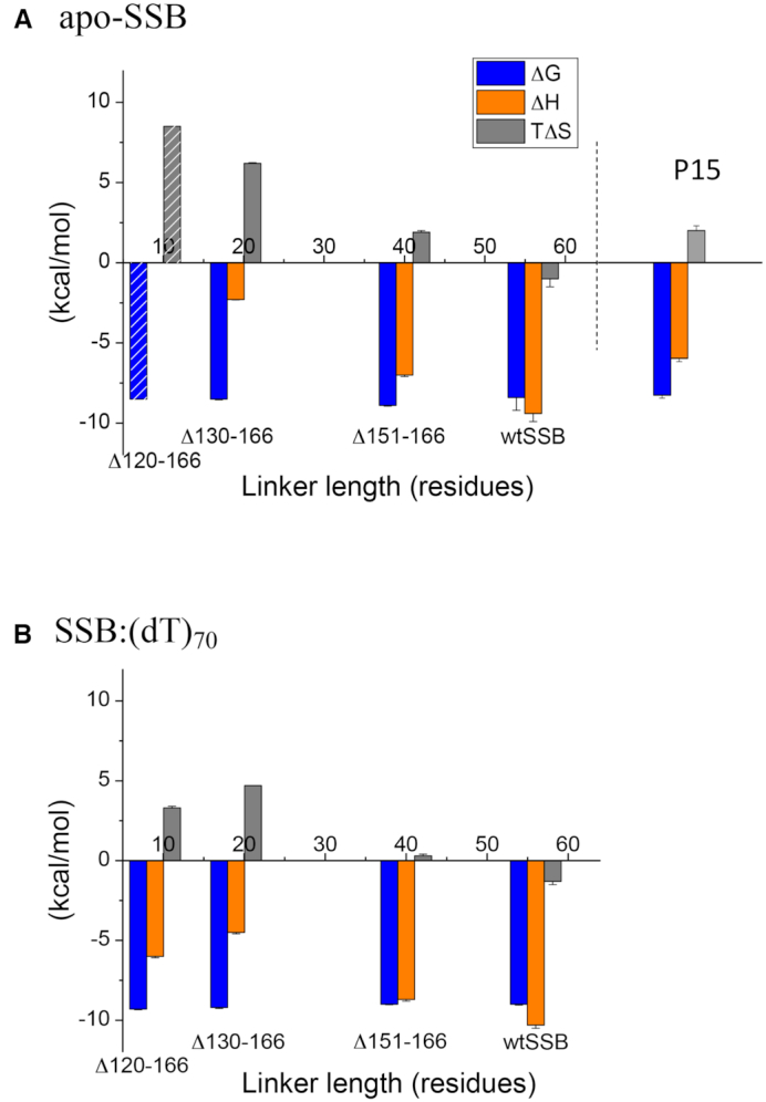
Escherichia coli single strand (ss) DNA binding (SSB) protein protects ssDNA intermediates and recruits at least 17 SSB interacting proteins (SIPs) during genome maintenance. The SSB C-termini contain a 9 residue acidic tip and a 56 residue intrinsically disordered linker (IDL). The acidic tip interacts with SIPs; however a recent proposal suggests that the IDL may also interact with SIPs. Here we examine the binding to four SIPs (RecO, PriC, PriA and χ subunit of DNA polymerase III) of three peptides containing the acidic tip and varying amounts of the IDL. Independent of IDL length, we find no differences in peptide binding to each individual SIP indicating that binding is due solely to the acidic tip. However, the tip shows specificity, with affinity decreasing in the order: RecO > PriA ∼ χ > PriC. Yet, RecO binding to the SSB tetramer and an SSB-ssDNA complex show significant thermodynamic differences compared to the peptides alone, suggesting that RecO interacts with another region of SSB, although not the IDL. SSB containing varying IDL deletions show different binding behavior, with the larger linker deletions inhibiting RecO binding, likely due to increased competition between the acidic tip interacting with DNA binding sites within SSB.


Figures

Figure 1.
Figure 1.
Structure of Escherichia coli SSB. (A) Model for the structure of an SSB tetramer complexed with (dT)70 in the (SSB)65 binding mode (1). The IDL (purple lines) and the nine amino acid acidic tips (red boxes), are shown schematically. (B) A cartoon representation of the domain structure of an SSB subunit showing the N-terminal DNA binding domain (DBD) (residues 1–112), the 56 amino acid IDL (residues 113–168) and the acidic tip (residues 169–177). (C) Sequences of the SSB-Ct peptides with the PXXP motifs underlined in black.
Figure 2.
Figure 2.
The acidic tip peptide, P15, binds with specificity to four SIPs. The results of ITC titrations of P15 peptide into four SIPs at 25°C, (A) RecO (B) PriA (C) χ in buffer BTP (pH 8.0, 50 mM NaCl) and (D) PriC in buffer BTP (pH 8.0, 10 mM NaCl). A titration of P15 with PriC at 50 mM NaCl showed no binding. Upper panels show the raw titration data, plotted as the heat signal (microcalories per second) versus time (minutes), obtained for 22 injections (12 μl each) of P15 (50 μM) into a SIP solution (1–2 μM). Lower panels show the integrated heat responses per injection, normalized to the moles of injected P15, after subtraction of the heats of dilution obtained from the blank titration of P15 into buffer (empty squares). The smooth curves represent the best fit of the data to an n-independent and identical site model. Binding parameters from the fits are indicated in each panel as well as in Table 1 and Supplementary Table S1 (N = stoichiometry, K = association equilibrium binding constant, ΔH = binding enthalpy).
Figure 3.
Figure 3.
RecO binds to the IDL peptides only via the acidic tip. (A) Panels i–iii show the results of ITC studies of the binding of P15, P31 and P65 peptides to RecO, respectively. The peptides (40–50 μM) were titrated into RecO (2 μM) in buffer BTP (pH 8.0, 50 mM NaCl) at 25°C. The smooth curves are simulations for a 1:1 binding model using the best fit binding parameters indicated in each panel (see also Table 1 and Supplementary Table S1). (B) Values of ΔG° (blue), ΔH (orange) and TΔS° (gray) obtained from ITC experiments for P15 (○), P31 (Δ) and P65 (□) peptides binding to RecO performed at the indicated temperatures. Solid lines show fits of the data with linear regression. The raw data are shown in Supplementary Figure S3.
Figure 4.
Figure 4.
RecO binding to P15, wtSSB and wtSSB–(dT)70 indicates that RecO interacts with more than the acidic tip. Thermodynamic parameters for RecO binding to (A)-P15 peptide, (B)-wtSSB per C-terminus and (C)-wtSSB–(dT)70 complex per C-terminus in buffer BTP (pH 7.0, 200 mM NaCl); ΔHobs (red squares), ΔG°obs (blue circles) and TΔS°obs (green triangles).
Figure 5.
Figure 5.
RecO binding to SSB tetramers with portions of the IDL deleted. (A) Schematics and sequences of the series of SSB variant tetramers with varying C-terminal IDL deletions, where the PXXP motifs are underlined. The amino acid residues deleted in each variant are denoted. The wtSSB DNA binding domain (DBD) is shown as an ellipse (blue). (B) Results of ITC experiments for RecO binding to tetramers of: (i)-wtSSB, (ii)-SSBΔ151-166, (iii)-SSBΔ130-166 and (iv)-SSBΔ120-166. SSB constructs (9–10 μM) were titrated into RecO (1 μM) in buffer BTP (pH 7.0, 200 mM NaCl) at 25°C. The smooth curves are simulations of Equations (1) and (2) using the best fit parameters determined from each titration. The ΔH for RecO binding per C-terminus is indicated on each panel. The binding parameters are shown in Table 3 and Supplementary Table S4. (C) The results of sedimentation velocity experiments, plotted as the c(s) distribution (70) for each SSB construct (0.35 μM tetramer) in the absence (black lines) and presence (red lines) of 2.5 molar excess of RecO (3.5 μM) per C-terminus. The sedimentation coefficients of each SSB are 1.4 S, 1.4 S, 1.3 S and 1.3 S, respectively, corresponding to the tetrameric species. Two peaks are observed in the presence of RecO. The peak at 0.8 S is due to free RecO. The SSB peaks are shifted to 1.7 S, 1.6 S and 1.5 S, and 1.6S, respectively, indicating binding of at least one RecO per SSB tetramer.
Figure 6.
Figure 6.
RecO binding to the linker deletion variants of SSB tetramer–(dT)70 complexes. Results of ITC experiments for RecO binding to complexes of (dT)70 bound to the SSB tetramers: (A)-wtSSB, (B)-SSBΔ151-166, (C)-SSBΔ130-166 and (D)-SSBΔ120-166. SSB–(dT)70 complexes (9–10 μM) were titrated into RecO (1 μM) in buffer BTP (pH 7.0, 200 mM NaCl) at 25°C. The smooth curves are simulations of Equations (1) and (2) using the best fit parameters determined from each titration. The ΔH for RecO binding per C-terminus is indicated on each panel. The binding parameters are shown in Table 3.
Figure 7.
Figure 7.
Thermodynamic parameters for the interactions of RecO with wtSSB and three SSB linker deletion variants. The thermodynamic parameters (per SSB C-terminus) obtained from the ITC experiments in Figures 5 and 6 (from Supplementary Table S4) are shown for binding of RecO to wtSSB, SSBΔ151-166, SSBΔ130-166 and SSBΔ120-166. (A) Values for RecO binding to apo-SSB tetramers; ΔG° (left-blue), ΔH (middle-orange), TΔS° (right-gray). Since ΔH is undetectable (∼0 kcal/mol) for SSBΔ120-166 binding to RecO, the indicated values of ΔG° and TΔS° were estimated by assuming that ΔG° is the same as for SSBΔ130-166. The parameters obtained for P15 binding to RecO are shown for comparison. (B) Values for RecO binding to SSB tetramers bound to (dT)70; ΔG° (left-blue), ΔH (middle-orange), TΔS° (right-gray).

Similar articles

Cited by

References

    1. Raghunathan S., Kozlov A.G., Lohman T.M., Waksman G.. Structure of the DNA binding domain of E. coli SSB bound to ssDNA. Nat. Struct. Biol. 2000; 7:648–652. - PubMed
    1. Chase J.W., Williams K.R.. Single-stranded DNA binding proteins required for DNA replication. Annu. Rev. Biochem. 1986; 55:103–136. - PubMed
    1. Meyer R.R., Laine P.S.. The single-stranded DNA-binding protein of Escherichia coli. Microbiol. Rev. 1990; 54:342–380. - PMC - PubMed
    1. Lohman T.M., Ferrari M.E.. Escherichia coli single-stranded DNA-binding protein: multiple DNA-binding modes and cooperativities. Annu. Rev. Biochem. 1994; 63:527–570. - PubMed
    1. Shereda R.D., Kozlov A.G., Lohman T.M., Cox M.M., Keck J.L.. SSB as an organizer/mobilizer of genome maintenance complexes. Crit. Rev. Biochem. Mol. Biol. 2008; 43:289–318. - PMC - PubMed

Publication types

MeSH terms

LinkOut - more resources