. 2019 Sep 10:309:231-243.
doi: 10.1016/j.jconrel.2019.07.025. Epub 2019 Jul 19.

Nanoparticulate delivery of potent microtubule inhibitor for metastatic melanoma treatment

Affiliations

Nanoparticulate delivery of potent microtubule inhibitor for metastatic melanoma treatment

Jitender Bariwal et al. J Control Release. .

Abstract

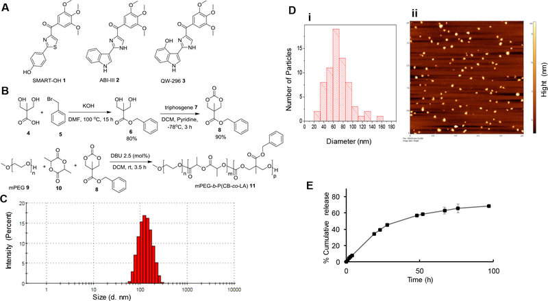
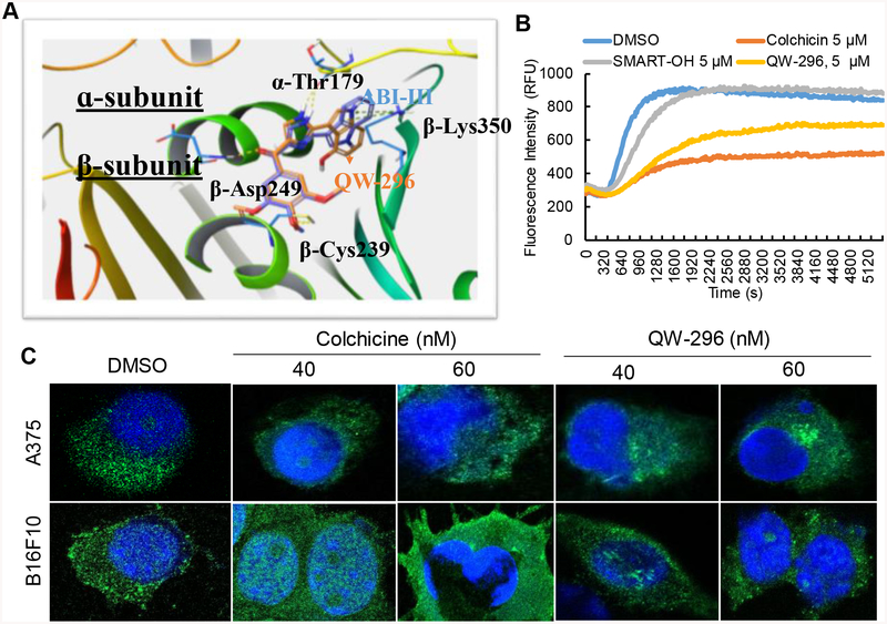
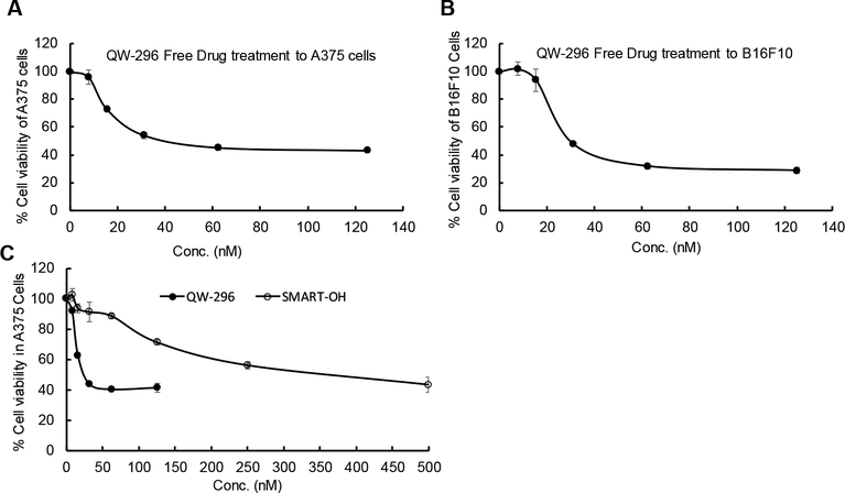
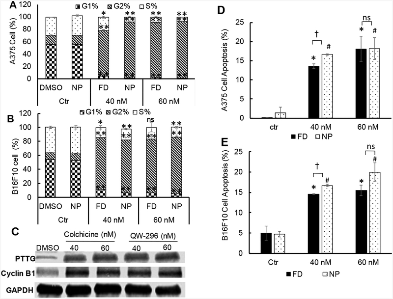
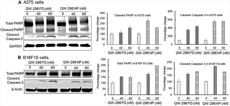
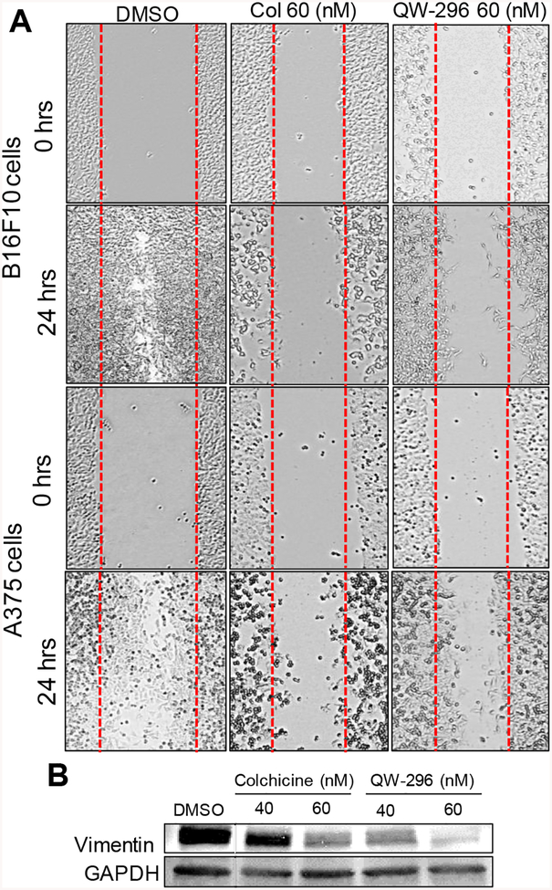
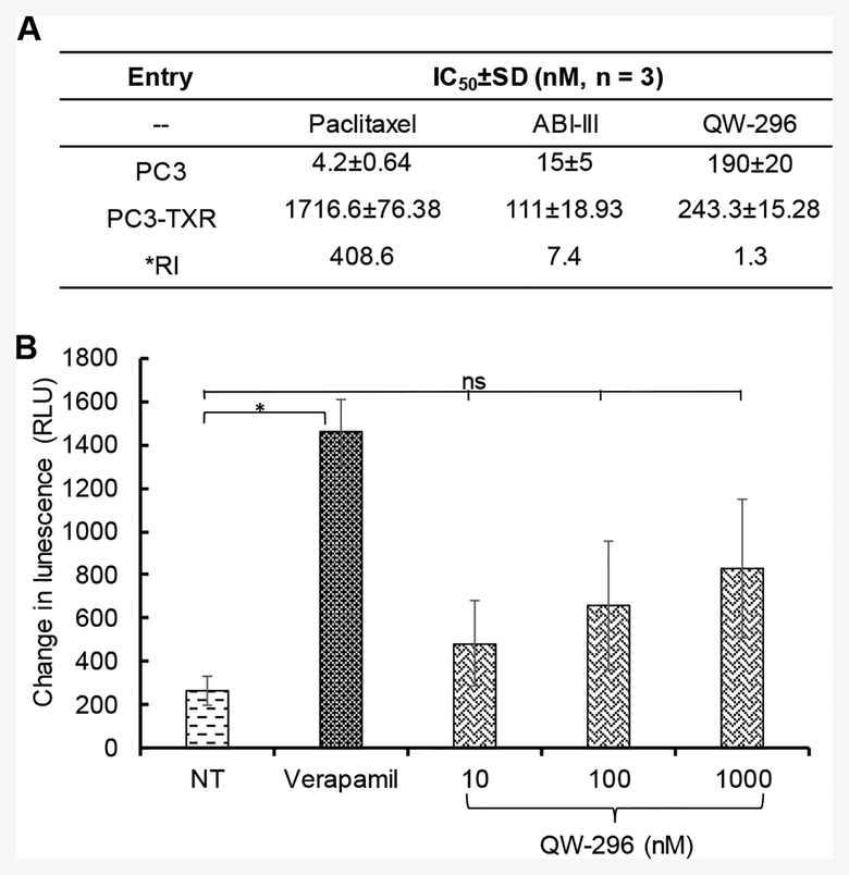
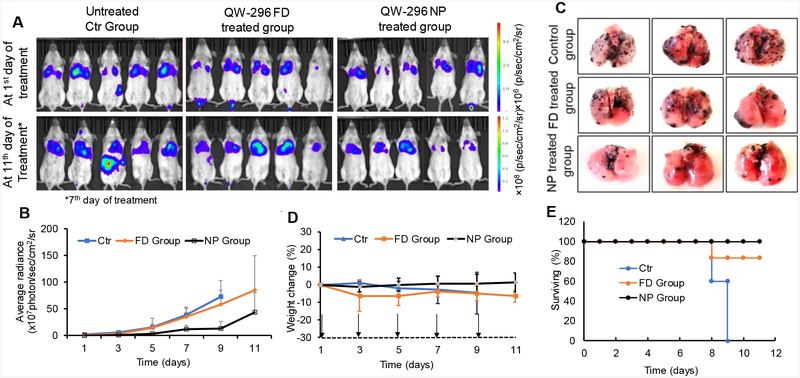
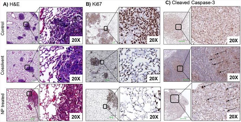
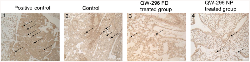
Melanoma is the most aggressive type of skin cancer, which readily metastasizes through lymph nodes to the lungs, liver, and brain. Since the repeated administration of most chemotherapeutic drugs develops chemoresistance and severe systemic toxicities, herein we synthesized 2-(4-hydroxy-1H-indol-3-yl)-1H-imidazol-4-yl)(3,4,5-trimethoxyphenyl) methanone (abbreviated as QW-296), a novel tubulin destabilizing agent with little susceptible to transporter-mediated drug resistance. QW-296 disturbed the microtubule dynamics at the nanomolar concentration in A375 and B16F10 melanoma cells. QW-296 binding to colchicine-binding site on tubulin protein was confirmed by molecular modeling and tubulin polymerization assay. QW-296 significantly inhibited A375 and B16F10 cell proliferation, induced G2/M cell cycle arrest and led to apoptosis and cell death. To improve its aqueous solubility, QW-296 was encapsulated into methoxy poly(ethyleneglycol)-b-poly(carbonate-co-lactide) [mPEG-b-P(CB-co-LA)] polymeric nanoparticles by solvent evaporation, with the mean particle size of 122.0 ± 2.28 nm and drug loading of 3.70% (w/w). Systemic administration of QW-296 loaded nanoparticles into C57/BL6 albino mice bearing lung metastatic melanoma at the dose of 20 mg/kg 4 times a week for 1.5 weeks resulted in significant tumor regression and prolonged mouse median survival without significant change in mouse body weight. In conclusion, QW-296 loaded nanoparticles have the potential to treat metastatic melanoma.

Keywords: Melanoma; Microtubule/tubulin inhibitor; Nanoparticles; Tubulin polymerization.


Conflict of interest statement

Conflicts of Interest

No potential conflicts of interest were disclosed.

Figures

Figure 1.
Figure 1.
Structure of colchicine inhibitors, mPEG-based polymer synthesis, and nanoparticle characterization. A) Chemical structure of SMART-OH, ABI-III and QW-296, B) Synthesis of mPEG-poly(carbonate-co-lactide). C) The particle size distribution of polymeric nanoparticles by w/o emulsification as determined by dynamic light scattering (DLS). D) Surface morphology and particle size distribution of nanoparticles by AFM. E) In vitro release of QW-296 from nanoparticles at pH 7.4. Drug concentrations were measured by HPLC. Results are expressed as the mean ± SD (n = 3).
Figure 2.
Figure 2.
Confirmation of the mechanism of action of QW-296. A) Proposed binding poses of ABI-III and QW-296 in the tubulin crystal structure (PDB code: 5H7O). Superposition of ABI-III (purple tube model; glide docking score −11.2) with QW-296 (orange tube model; glide docking score −11.9). B) Effect of QW-296 on tubulin polymerization in in vitro. C) Confocal images of A375 and B16F10 melanoma cells exposed to colchicine and QW-296 or the vehicle (DMSO) for 24 h.
Figure 3.
Figure 3.
Cell viability assay using QW-296 in melanoma cells. A) Cytotoxicity of QW-296 in A375 cells, B) in B16F10 cells and C) comparison of cytotoxicity of QW-296 and SMART-OH in A375 cells after 48 h of treatment.
Figure 4.
Figure 4.
Effect of QW-296 on cell cycle analysis and induction of apoptosis in melanoma cells. A) Cell cycle analysis after treatment with QW-296 (free drug and loaded nanoparticles) to A375 cells, and B) B16F10 cells. C) Effect of the QW-296 treatment on the expression of G2/M related proteins PTTG and Cyclin B1. D) Induction of apoptosis by QW-296 as free drug and loaded in nanoparticles in A375 cells and E) B16F10 cells after 24 h of incubation. *, p<0.05 (comparison between control vs free drug), #, p<0.05 (comparison between control vs NP), †, p<0.05 (comparison between free QW-296 vs QW-296 loaded NP), ns, p>0.05.
Figure 5.
Figure 5.
Estimation of apoptotic protein expression (PARP and Cleaved Caspase 3) by WB in A) A375 cells and B) B16F10 cells after treatment with QW-296 in cosolvent and QW-296 loaded nanoparticles after 48 h of treatment.
Figure 6.
Figure 6.
Effect of QW-296 and colchicine on cell migration and EMT marker. A) Scratch assay in B16F10 and A375 cells. B) Dose-dependent decrease in vimentin in A375 cells.
Figure 7.
Figure 7.
Determination of resistance index and possible mechanism to overcome chemoresistance by QW-296. A) Anti-proliferative activity of paclitaxel (PTX), ABI-III and QW-296 on paclitaxel-resistant PC3-TXR cells and non-resistant PC3 cells. B) Effect of QW-296 on Pgp ATPase activity. Change in luminescence compared to 100 μM Na3VO4 treated samples was plotted (mean ± SD, n = 3). QW-296 at lower concentrations (10 and 100 nM) showed a non-significant effect compared to NT control on Pgp ATPase activity. *, p < 0.05.
Figure 8.
Figure 8.
In vivo therapeutic effect of QW-296 as FD and QW-296 loaded nanoparticles (NPs) in lung tumor metastasis mice model. A) In vivo representative bioluminescent images at day 1 and day 11 of treatment. Bioluminescent images of mice from control (blank NP), QW-296 FD group and QW-296 loaded in NP groups were taken every alternate day during the treatment (n = 6), *indicates bioluminescent images of control group on day 7th of the study. B) Radiance intensity plot of all three treatment groups measured from day 1 of treatment to the end of the study. C) Representative lungs showing tumor nodules of each group excised after sacrificing the mice at the end of the study. D) Mouse % body weight change during treatment for all the three groups. E) Survival plot of all the animals used using QW-296 in cosolvent and QW-296 loaded in NPs. Data represented as the mean ± SD (n=6). Arrow indicates the day of injection.
Figure 9.
Figure 9.
Effect of QW-296 treatment on tumor growth and tumor cell apoptosis. Lung tumor nodules from control, QW-296 in cosolvent and QW-296 loaded nanoparticles (NPs) treated groups were stained by A) hematoxylin and eosin (H&E), B) proliferation marker Ki67, and C) apoptosis marker cleaved Caspase 3.
Figure 10.
Figure 10.
DeadEnd™ colorimetric TUNEL assay of mouse lung tissues to indicate DNA fragmentation in 1) positive control, 2) control group, 3) mice treated with 20mg/kg QW-296 in cosolvent system, and 4) mice treated with 20 mg/kg equivalent dose of QW-296 loaded NPs. Slides were observed under the light microscope and arrows show dark stained nuclei which indicate DNA fragmentation and nuclear condensation.

References

    1. Kaur R, Kaur G, Gill RK, Soni R, Bariwal J. Recent developments in tubulin polymerization inhibitors: An overview. Eur J Med Chem. 2014;87:89–124. - PubMed
    1. Li CM, Chen J, Lu Y, et al. Pharmacokinetic optimization of 4-substituted methoxybenzoyl-aryl-thiazole and 2-aryl-4-benzoyl-imidazole for improving oral bioavailability. Drug Metab Dispos. 2011;39(10):1833–1839. - PMC - PubMed
    1. Arnst KE, Wang Y, Hwang DJ, et al. A potent, metabolically stable tubulin inhibitor targets the colchicine binding site and overcomes taxane resistance. Cancer Res. 2018;78(1):265–277. - PubMed
    1. Gigant B, Wang C, Ravelli RB, et al. Structural basis for the regulation of tubulin by vinblastine. Nature. 2005;435(7041):519–522. - PubMed
    1. Dorleans A, Gigant B, Ravelli RB, Mailliet P, Mikol V, Knossow M. Variations in the colchicine-binding domain provide insight into the structural switch of tubulin. Proc Natl Acad Sci U S A. 2009;106(33):13775–13779. - PMC - PubMed

Publication types

MeSH terms