. 2019 Aug;572(7769):387-391.
doi: 10.1038/s41586-019-1439-1. Epub 2019 Jul 22.

Regulation of phosphoribosyl ubiquitination by a calmodulin-dependent glutamylase

Affiliations

Regulation of phosphoribosyl ubiquitination by a calmodulin-dependent glutamylase

Ninghai Gan et al. Nature. 2019 Aug.

Abstract

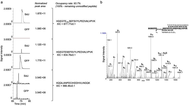
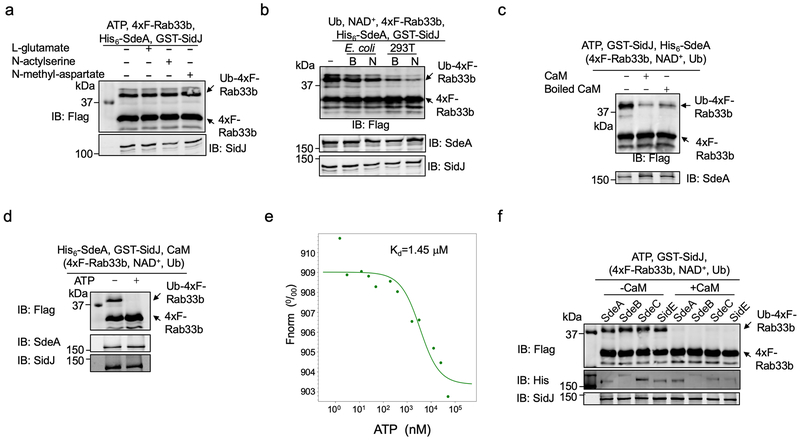
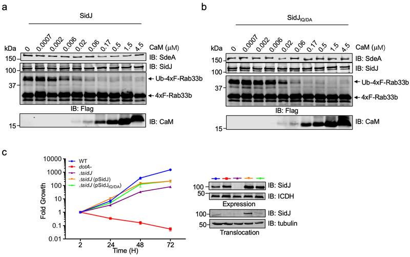
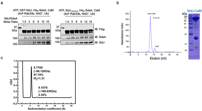
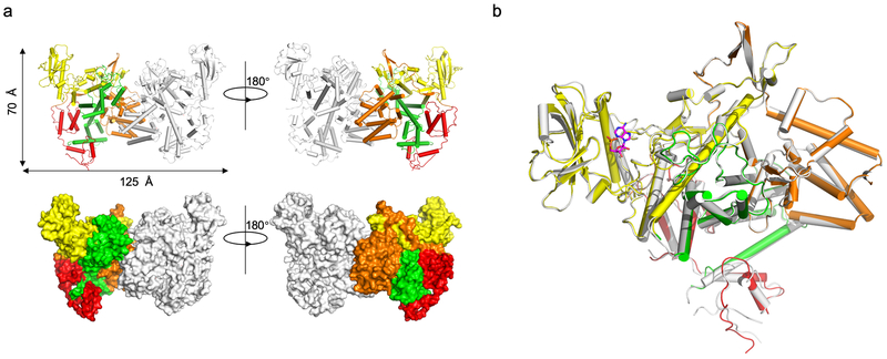
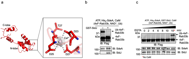
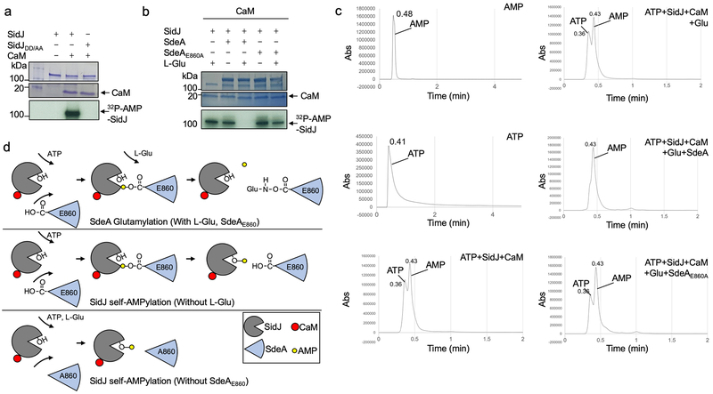
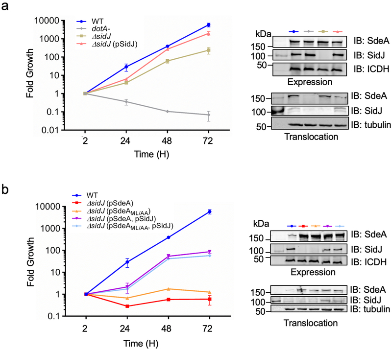
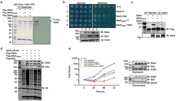
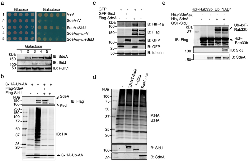
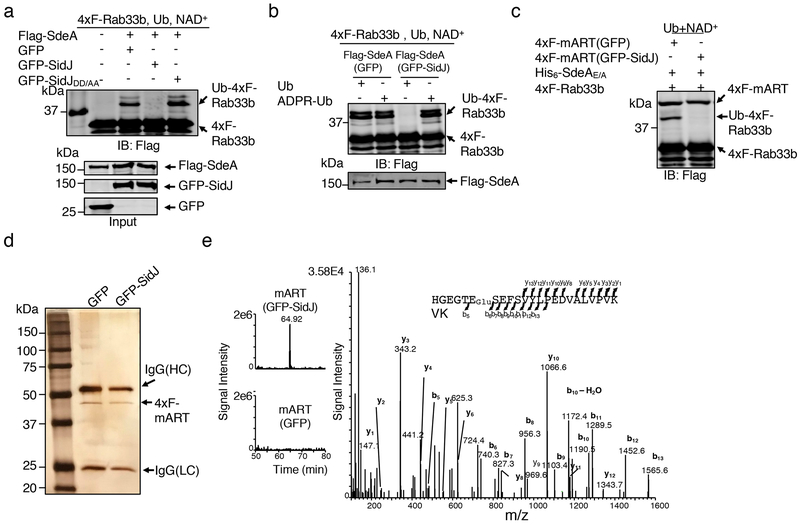
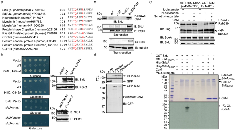
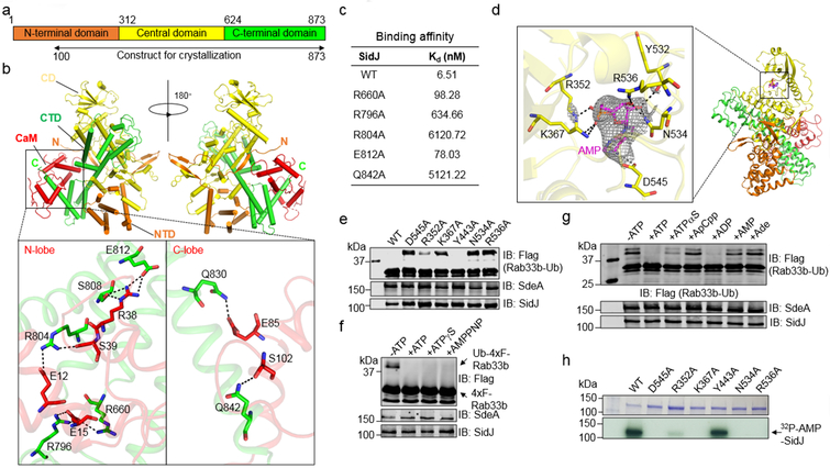
The bacterial pathogen Legionella pneumophila creates an intracellular niche permissive for its replication by extensively modulating host-cell functions using hundreds of effector proteins delivered by its Dot/Icm secretion system1. Among these, members of the SidE family (SidEs) regulate several cellular processes through a unique phosphoribosyl ubiquitination mechanism that bypasses the canonical ubiquitination machinery2-4. The activity of SidEs is regulated by another Dot/Icm effector known as SidJ5; however, the mechanism of this regulation is not completely understood6,7. Here we demonstrate that SidJ inhibits the activity of SidEs by inducing the covalent attachment of glutamate moieties to SdeA-a member of the SidE family-at E860, one of the catalytic residues that is required for the mono-ADP-ribosyltransferase activity involved in ubiquitin activation2. This inhibition by SidJ is spatially restricted in host cells because its activity requires the eukaryote-specific protein calmodulin (CaM). We solved a structure of SidJ-CaM in complex with AMP and found that the ATP used in this reaction is cleaved at the α-phosphate position by SidJ, which-in the absence of glutamate or modifiable SdeA-undergoes self-AMPylation. Our results reveal a mechanism of regulation in bacterial pathogenicity in which a glutamylation reaction that inhibits the activity of virulence factors is activated by host-factor-dependent acyl-adenylation.


Figures

Extended Data Fig. 1
Extended Data Fig. 1. Determination of the modification rate on E860 of SdeA.
a. Peak areas of the extracted-ion chromatograms (XIC) were normalized based on the area of the unmodified peptide -I608IQQILANPDCIHDDHVLINGQK630−. The occupancy rate of glutamylation on residue was calculated based on the consumption of the unmodified -H855GEGTESEFSVYLPEDVALVPVK877− in samples from cells cotransfected to express GFP-SidJ compared to those of controls from cells transfected to express GFP. b. SidJ induces a 258.09 Dalton post-translation modification on E860 within the mART motif of SdeA. 4xFlag-mART purified from HEK293T cells coexpressing SidJ detected by silver staining (Fig. 2d) was analyzed by mass spectrometric analysis. Tandem mass (MS/MS) spectrum shows the fragmentation profile of the modified peptide -H855GEGTEGluGluSEFSVYLPEDVALVPVK877−, including ions b5 and b6 that confirms the modification site at the E860 residue. In each case, similar results were obtained in three independent experiments.
Extended Data Fig. 2
Extended Data Fig. 2. The effects of cell lysates, ATP, heat treatment on CaM on the activity of SidJ and its inhibition of the activity of all members of the SidE family.
a. Inhibition of SdeA activity does not occur in in vitro reactions containing L-glutamate or each of its two structural isomers. L-glutamate, N-acetylserine or N-methyl-aspartate was incubated with SdeA, SidJ and ATP for 2 h before assaying for the activity of SdeA. b. A molecule(s) from mammalian cells is required for SidJ to inhibit SdeA. Lysates from E. coli or HEK293T cells were added to reactions containing SdeA and SidJ for 2 h before measuring the activity of SdeA. c. Heat treatment does not completely abolish CaM activity. CaM or CaM treated by heating at 100°C for 5 min was included in reactions that allow glutamylation of SdeA for 2 h. A cocktail containing 4xF-Rab33b, NAD+ and ubiquitin was added to each reaction. Samples were resolved by SDS-PAGE and detected for Rab33b ubiquitination after another 2 h incubation at 37°C. d. The activity of SidJ requires ATP. His6-SdeA was incubated with GST-SidJ, L-glutamate and CaM in reactions with or without 1 mM ATP for 2 h, 4xF-Rab33b, NAD+ and ubiquitin were added to each reaction. After another 2 h incubation, the activity of SdeA was evaluated by the production of ubiquitinated Ra33b. Protein components in the reactions were detected by immunoblotting with specific antibodies. e. The binding of ATP by SidJ. Binding of ATP by purified SidJ was evaluated using Microscale thermophoresis in which the concentration of SidJ was kept constant. The dissociation constant (Kd) was determined by the NanoTemper Analysis 2.2.4 software. f. SidJ inhibits the activity of members of the SidE family. Recombinant protein of each of SidE family protein was incubated with ATP, L-glutamate and GST-SidJ in the presence or absence of CaM for 2 h, a cocktail containing 4xF-Rab33b, NAD+ and ubiquitin was added to the reactions. After additional 2 h incubation, modification of Rab33b was detected by immunoblotting with a Flag-specific antibody. The formation of Ub-4xF-Rab33b is indicated by a shift in molecular weight. In each panel, data shown were one representative from at least three independent experiments that had similar results.
Extended Data Fig. 3
Extended Data Fig. 3. The IQ motif of SidJ is required for its optimal response to CaM
a-b. The IQ motif is required for the optimal activity of SidJ in response to CaM. Serially diluted CaM were preincubated with SidJ (a) or the SidJIQ mutant (b) and SdeA in the glutamylation buffer at 37°C for 2 h. A cocktail containing 4xFlag-Rab33b, NAD+, and ubiquitin was added to the reactions. After incubation for another 2 h at 37°C. Proteins separated by SDS-PAGE were probed with the indicated antibodies. SidJIQ/DA, I841, Q842 were mutated to Asp and Ala, respectively. In each panel, data shown were one representative from at least three independent experiments that had similar results. c. The SidJIQ mutant complements the intracellular growth defect of the ΔsidJ mutant. Acanthamoeba castellanii was infected with the indicated bacterial strains and intracellular bacteria were determined at the indicated time points. Each strain was done in triplicate and similar results were obtained in two independent experiments. Results are from one representative experiment done in triplicate from three independent experiments; error bars represent s.e.m. (n = 3).
Extended Data Fig. 4
Extended Data Fig. 4. SidJ forms stable heterodimer with CaM at the molar ratio of 1:1
a. SidJ∆N99 maintains the ability to inhibit SdeA activity at levels comparable to full-length SidJ. SdeA was incubated with GST-SidJ or SidJ∆N99 at indicated molar ratios in reactions containing ATP, L-Glu, CaM for 2 h at 37°C. A cocktail containing 4xFlag-Rab33b, NAD+, and ubiquitin was added to each reaction for additional 2 h at 37°C, proteins resolved by SDS-PAGE were probed with the indicated antibodies. SdeA activity was measured by the production of ubiquitinated Rab33b as indicated by a shift in molecular weight. b. Size exclusion chromatography profiles of SidJ-CaM. Purified proteins were separated by a Superdex 200 increase 10/300 column (GE Healthcare) on an AKTA pure system (left). Fractions with strong absorbance at OD260 were collected and analyzed by SDS-PAGE followed by detection with Coomassie brilliant blue staining (right). c. The heterodimer formed between SidJ∆N99 with CaM is a monomer. Analytical ultracentrifugation analysis yielded a sedimentation coefficient of 5.770 S, and a molecular mass of approximately 96.12 kDa, indicating the heterodimer of SidJ∆N99 and CaM. In each panel, data shown were one representative from at least three independent experiments that had similar results.
Extended Data Fig. 5
Extended Data Fig. 5. Overall structure of SidJ-CaM complex in one asymmetric unit and the comparison of complex structures with or without AMP
a. Two views of the structure of the SidJ-CaM heterodimer in the asymmetric unit (ASU) displayed as ribbon diagram (upper panel) and surface rendering (lower panel), one of the SidJ-CaM heterodimer is colored as shown in Fig. 4 and the other one is colored in grey. b. Superimposition of the structures of the SidJ-CaM and SidJ-CaM-AMP. The SidJ-CaM-AMP ternary complex is colored as shown in Fig. 4d and SidJ-CaM binary complex is colored in grey.
Extended Data Fig. 6
Extended Data Fig. 6. Interactions between CaM and Ca2+ from the crystal structures and the role of Ca2+ on the activation of SidJ by CaM
a. Key residues of CaM involved in the interaction with Ca2+. Ca2+ is coordinated by D21, D23, D25 and T27 of CaM are shown in red sticks. Ca2+ is shown in pink sphere. Electron density of an SA Fo-Fc omit map for Ca2+ contoured at 3.0 σ. b. Dialysis against 20 mM EGTA does not abolish the activity of SidJ. All proteins used in the reactions were dialyzed against a buffer containing 20 mM EGTA for 14 h. SdeA was incubated with SidJ in reactions containing ATP and dialyzed CaM for 2 h at 37°C. Reactions without SidJ were established as a control. A cocktail containing 4xFlag-Rab33b, NAD+, and ubiquitin was added to each reaction. After further incubation for 2 h at 37°C, proteins resolved by SDS-PAGE were probed with the indicated antibodies. SdeA activity was measured by the production of ubiquitinated Rab33b as indicated by a shift in molecular weight. c. The activity of SidJ is not sensitive to 10 mM EGTA. SdeA was first incubated with SidJ for glutamylation with indicated amounts of EGTA for 2 h at 37°C. NAD+, 4xFlag-Rab33b and ubiquitin were then supplemented to the reactions, which were allowed to proceed for 2 h at 37°C before being resolved by SDS-PAGE. Rab33b modification was detected as described in b. Proteins in the reactions were detected by immunoblotting with specific antibodies. In panels b-c, similar results were obtained in at least three independent experiments.
Extended Data Fig. 7
Extended Data Fig. 7. The mechanism of SidJ induced CaM dependent self-AMPlyation and SdeA glutamylation
a. SidJ induces self-AMPylation in a CaM dependent manner. SidJ was incubated with 32P-α-ATP, Mg2+, with or without CaM for 2 h at 37°C. After separation by SDS-PAGE, the incorporation of 32P-α-ATP was detected by autoradiography. b. SdeA glutamylation by SidJ interferes with SidJ self-AMPylation. SidJ was incubated with 32P-α-ATP, Mg2+, CaM for 2 h at 37°C. When needed L-Glu, SdeA, SdeAE860A were supplemented. After separation by SDS-PAGE, the incorporation of 32P-α-ATP was detected by autoradiography. c. SdeA glutamylation by SidJ accelerates ATP hydrolysis and AMP release. SidJ was incubated with indicated components for 2 h at 37°C. Samples were analyzed by HPLC. AMP and ATP were used as standard. In panels a-c, data shown were one representative from at least three independent experiments that had similar results. d. Schematic model of SidJ induced glutamylation and AMPylation. SidJ incudes glutamylation on SdeAE860 when ATP and L-Glu are supplemented in reaction. In reactions missing L-Glu or modifiable SdeA, SidJ induces self-AMPylation.
Extended Data Fig. 8
Extended Data Fig. 8. Intracellular growth phenotypes associated the sidJ mutant expressing SdeA and its mutants.
a. Intracellular defects of the L. pneumophilasidJ mutant can be complemented by SidJ expressed from a multicopy plasmid. The indicated strains were used to infect A. castellanii at an MOI of 0.05 and the growth of the bacteria was evaluated at 24 h intervals. Fold growth was calculated based on total bacterial counts at the indicated time points and those of the 2 h point. b. Overexpression of a SdeA mutant defective in substrate recognition inhibits intracellular growth of the ∆sidJ mutant. Intracellular growth of the indicated L. pneumophila strains in A. castellanii was evaluated as described in a. In each panel, the expression of SidJ, SdeA and its mutants in bacterial cells and their translocation into infected cells was determined by immunoblotting from total bacterial cell lysates and the saponin-soluble fraction of infected cells, with ICDH and tubulin as loading controls, respectively (right panels). In each case, results are from one representative experiment done in triplicate from three independent experiments; error bars represent s.e.m. (n = 3).
Extended data Fig. 9
Extended data Fig. 9. SidJ functions to regulate the activity of SdeA during L. pneumophila infection
a. SdeAE860D is resistant to glutamylation catalyzed by SidJ. SdeA, SdeAE860A or SdeAE860D was added to reactions containing GST-SidJ, 14C-glutamate ATP and CaM and the reactions were allowed to proceed for 2 h at 37°C. After separation by SDS-PAGE, the incorporation of 14C-glutamate was detected by autoradiography. b. Yeast toxicity induced by SdeAE860D cannot be suppressed by SidJ. A plasmid that directs the expression of SidJ was introduced into yeast strains expressing SdeA or SdeAE860D from a galactose inducible promoter, serially diluted yeast cells were spotted onto glucose or galactose medium for 2 d and the growth of the cells was evaluated by imaging (upper panels). The expression of SidJ, SdeA and SdeAE860D was determined by immunoblotting with specific antibodies. The PGK1 (3-phosphoglyceric phosphokinase-1) was probed as a loading control (lower panels). c. SdeAE860D still ubiquitinates Rab33b. Reactions containing the indicated components were allowed to proceed for 2 h at 37°C, samples were then resolved by SDS-PAGE and ubiquitination of Rab33b was probed by immunoblotting with a Flag-specific antibody to detect the production of modified Rab33b with a higher molecular weight. d. SdeAE860D-mediated protein ubiquitination in mammalian cells is insensitive to SidJ. HEK293T cells were transfected to express the indicated proteins for 16-18 h. Cleared cell lysates were subjected to SDS-PAGE and immunoblotting with an HA specific antibody to detected proteins ubiquitinated by 3xHA-Ub-AA. The levels of SdeA, SdeAE860D and SidJ were assessed by antibodies specific for these proteins. Note that coexpression of SidJ reduced the ubiquitination induced by SdeA but not SdeAE860D. In panels a-d, data shown were one representative from at least three independent experiments that had similar results. e. The effects of SidJ on intracellular growth defect caused by overexpression of SdeA or SdeAE860D. The indicated L. pneumophila strains were used to infect Acanthamoeba castellanii at an MOI of 0.05 and the growth of the bacteria was evaluated at 24 h intervals. Fold growth was calculated based on total bacterial counts at the indicated time points. Note that the difference between strain ∆sidJ (pSdeA) and ∆sidJ (pSdeA, pSidJ). The growth defect caused by overexpressing the SdeAE860D mutant cannot be rescued by SidJ. The levels of relevant proteins in bacterial cells and in infected cells were probed by immunoblotting from total bacterial cell lysates and the saponin-soluble fraction of infected cells, with ICDH and tubulin as loading controls, respectively (right panels). Results showen are from one representative experiment done in triplicate from three independent experiments; error bars represent s.e.m. (n = 3).
Fig. 1
Fig. 1. SidJ antagonizes the effects of SdeA in eukaryotic cells
a. SidJ suppresses the yeast toxicity of SdeAH277A. Diluted cells from yeast strains inducible expressing SdeA or SdeAH277A that harbor the vector or a SidJ construct were spotted onto the indicated media and grew for 2 d (top). The expression of relevant proteins was probed by immunoblotting (bottom). The 3-phosphoglyceric phosphokinase-1 (PGK1) was probed as a loading control. V, vector. b. SidJ abrogates SdeA-mediated ubiquitination in mammalian cells. Lysates of HEK293T cells expressing the indicated proteins were detected by immunoblotting with an HA-specific antibody to detect 3xHA-Ub-AA and proteins modified by 3xHA-Ub-AA. The expression of Flag-SdeA and Flag-SidJ was also probed. c. SidJ rescues HIF-1α degradation blocked by SdeA. Lysates of HEK293T cells expressing the indicated proteins were resolved by SDS-PAGE and probed with antibodies specific for the epitope tags or relevant proteins. d. SidJ from E. coli or HEK293T cells cannot deubiquitinate proteins modified by SdeA. Proteins modified by 3xHA-Ub-AA obtained by immunoprecipitation were treated with GST-SidJ from E. coli, Flag-SidJ from HEK293T or SdeA1-193. Note that none of these proteins caused a reduction in the ubiquitination signals. e. GST-SidJ does not inhibit SdeA-induced ubiquitination in vitro. SidJ was co-incubated with SdeA for 2 h at 37°C and SdeA activity was assayed. A Flag-specific antibody was used to detect modified and unmodified 4xFlag-Rab33b, judging by a shift in its molecular weight. SdeA and SidJ were probed with specific antibodies. SdeAE/A is a SdeA mutant defective in the mART activity carrying E860A and E862A mutations. The experiment in each panel was performed independently for at least 3 times with similar results.
Fig. 2
Fig. 2. SidJ post translationally modifies SdeA in mammalian cells and inhibits its activity to catalyze the production of ADP-ribosylated ubiquitin
a. Flag-SdeA coexpressed with SidJ fails to modify Rab33b. Flag-SdeA from HEK293T cells coexpressing relevant proteins was used to ubiquitinate 4xFlag-Rab33b. Ub-Rab33b was detected as described in Fig. 1. SidJDD/AA is a SidJ mutant defective in suppressing the yeast toxicity of SdeA that carries D542A and D545A mutations. b. Flag-SdeA coexpressed with SidJ retains the ability to ubiquitinate Rab33b with ADPR-Ub. ADPR-Ub or ubiquitin was incubated with Flag-SdeA purified from HEK293T cells coexpressing GFP or GFP-SidJ. NAD+ was included in reactions receiving ubiquitin. Rab33b modification was detected with a Flag-specific antibody. c. SidJ attacks the mART activity of SdeA. 4xFlag-mART (SdeA563-910) purified from HEK293T cells coexpressing GFP or GFP-SidJ was incubated with 4xFlag-Rab33b, ubiquitin, NAD+ and His6-SdeAE/A for 2 h at 37°C before ubiquitination detection. d-e. SidJ induces a 129.04 Dalton post-translational modification on E860 of SdeA. Mass spectrometric analysis of 4xFlag-mART* (d) identified a posttranslational modification in the fragment -H855GEGTESEFSVYLPEDVALVPVK877− (e, left panel). Tandem mass (MS/MS) spectrum shows the fragmentation profile of the modified peptide -H855GEGTEGluSEFSVYLPEDVALVPVK877−, including ions b5 and b6 that confirm the modification site at E860 (e, right panel). The experiment in each panel was repeated three times with similar results.
Fig. 3
Fig. 3. Calmodulin is the host cofactor required for the glutamylase activity of SidJ
a. SidJ harbors an IQ motif. Alignment of the IQ domain of SidJ and that of several CaM-binding proteins. Conserved residues were highlighted in red. The accession number for each protein was included. b. The cmd1 gene suppresses the yeast toxicity of SidJ. Top two panels: Images of serially diluted yeast cells inducibly expressing sidJ or its IQ mutant spotted onto the indicated media for 2 d. Lower two panels: The suppression of SidJ toxicity by cmd1. The expression of SidJ in each strain was examined and PGK1 was probed as a loading control (right panels). c-d. The interactions between SidJ and CaM. Beads coated with CaM were incubated with lysates of macrophages infected with the indicated bacterial strains to probe its binding to SidJ (c, top). SidJ in bacteria (c, middle) or translocated into the host cytosol (c, lower panel) was also examined. The bacterial isocitrate dehydrogenase (ICDH) and tubulin were probed as loading controls, respectively. Lysates of HEK293T cells transfected to express GFP-SidJ or GFP-SidJIQ/DA were incubated with CaM-coated beads (d). SidJ or SidJIQ/DA bound to CaM was probed by immunoblotting (lower panel). TCL, total cell lysates. e. Inhibition of SdeA activity by SidJ requires glutamate and CaM. CaM was added to a subset of a series of reactions containing SdeA, GST-SidJ and L-glutamate, N-acetylserine or N-methyl-aspartate. The activity of SdeA was measured by Rab33b ubiquitination. f. SidJ is a CaM-dependent glutamylase that modifies SdeA at E860. A series of reactions containing the indicated proteins, 14C-glutamate and ATP were allowed to proceed for 2 h at 37°C. The incorporation of 14C-glutamate was detected by autoradiography. Data shown in panels b-f were one representative from at least three experiments with similar results.
Fig. 4
Fig. 4. Structural analysis of the mechanism of SidJ-catalyzed glutamylation
a. Domain organization of SidJ. SidJ consists of the N-terminal domain (orange), the Central domain (yellow) and the C-terminal domain (green). b. Ribbon diagram representation of the SidJ-CaM complex. In top panels, the N-terminal domain (orange), Central domain (yellow), C-terminal domain (green) of SidJ and CaM (red) are shown. The view on top right is generated by rotating the structure shown on the top left by 180° around the indicated axis. The N and C termini of SidJ are labeled with letters. The missing residues are shown in dashed lines. Lower panels depict interactions between SidJ and the N-lobe/C-lobe of CaM. Residues important for binding are shown in sticks and hydrogen bonds are indicated by dashed lines. c. Binding of CaM to SidJ and its mutants. The binding affinity was evaluated using microscale thermophoresis. The dissociation constant (Kd) was calculated by the NanoTemper Analysis 2.2.4 software. Data shown were one representative from three experiments with similar results. d. Ribbon representation of the SidJ-CaM-AMP complex. Key residues of SidJ involved in AMP binding are shown in yellow sticks, AMP is labeled in magenta sticks. Hydrogen bonds are shown as dashed lines. Electron density of an SA (simulated annealing) Fo-Fc omit map for AMP contoured at 3.0 σ. e. Mutational analysis of residues important for binding AMP. Each SidJ mutant was incubated with SdeA, ATP, L-glutamate and CaM for 2 h before measuring SdeA’s ubiquitin ligase activity. f-g. Activation of SidJ by ATP analogs. The indicated compounds were incubated with SdeA, GST-SidJ, L-glutamate and CaM for 2 h at 37°C before monitoring SdeA’s activity in ubiquitinating Rab33b. Note that analogs defective in hydrolysis at the α site cannot activate SidJ. h. The role of residues important for AMP binding in SidJ self-AMPylation. Each SidJ mutant was incubated with 32P-α-ATP, Mg2+ and CaM for 2 h at 37°C and the incorporation of 32P-α-ATP was detected by autoradiography. In panels c, e-h, data shown were one representative from at least three independent experiments with similar results.

Comment in

References

    1. Qiu J & Luo ZQ Legionella and Coxiella effectors: strength in diversity and activity. Nat Rev Microbiol 15, 591–605, doi: 10.1038/nrmicro.2017.67 (2017). - DOI - PubMed
    1. Qiu J et al. Ubiquitination independent of E1 and E2 enzymes by bacterial effectors. Nature 533, 120–124, doi: 10.1038/nature17657 (2016). - DOI - PMC - PubMed
    1. Bhogaraju S et al. Phosphoribosylation of Ubiquitin Promotes Serine Ubiquitination and Impairs Conventional Ubiquitination. Cell 167, 1636–1649.e1613, doi: 10.1016/j.cell.2016.11.019 (2016). - DOI - PubMed
    1. Kotewicz KM et al. A Single Legionella Effector Catalyzes a Multistep Ubiquitination Pathway to Rearrange Tubular Endoplasmic Reticulum for Replication. Cell Host Microbe 21, 169–181, doi: 10.1016/j.chom.2016.12.007 (2017). - DOI - PMC - PubMed
    1. Liu Y & Luo ZQ The Legionella pneumophila effector SidJ is required for efficient recruitment of endoplasmic reticulum proteins to the bacterial phagosome. Infect Immun 75, 592–603 (2007). - PMC - PubMed

Publication types

MeSH terms

LinkOut - more resources