. 2019 Jul 21;8(7):1070.
doi: 10.3390/jcm8071070.

ATP Synthase Subunit Epsilon Overexpression Promotes Metastasis by Modulating AMPK Signaling to Induce Epithelial-to-Mesenchymal Transition and Is a Poor Prognostic Marker in Colorectal Cancer Patients

Affiliations

ATP Synthase Subunit Epsilon Overexpression Promotes Metastasis by Modulating AMPK Signaling to Induce Epithelial-to-Mesenchymal Transition and Is a Poor Prognostic Marker in Colorectal Cancer Patients

Yan-Jiun Huang et al. J Clin Med. .

Abstract

Metastasis remains the major cause of death from colon cancer. We intend to identify differentially expressed genes that are associated with the metastatic process and prognosis in colon cancer. ATP synthase epsilon subunit (ATP5E) gene was found to encode the mitochondrial F0F1 ATP synthase subunit epsilon that was overexpressed in tumor cells compared to their normal counterparts, while other genes encoding the ATP synthase subunit were repressed in public microarray datasets. CRC cells in which ATP5E was silenced showed markedly reduced invasive and migratory abilities. ATP5E inhibition significantly reduced the incidence of distant metastasis in a mouse xenograft model. Mechanistically, increased ATP5E expression resulted in a prominent reduction in E-cadherin and an increase in Snail expression. Our data also showed that an elevated ATP5E level in metastatic colon cancer samples was significantly associated with the AMPK-AKT-hypoxia-inducible factor-1α (HIF1α) signaling axis; silencing ATP5E led to the degradation of HIF1α under hypoxia through AMPK-AKT signaling. Our findings suggest that elevated ATP5E expression could serve as a marker of distant metastasis and a poor prognosis in colon cancer, and ATP5E functions via modulating AMPK-AKT-HIF1α signaling.

Keywords: AMPK; ATP5E; Colorectal Cancer; EMT; Metastasis.


Conflict of interest statement

The authors declare no conflict of interest.

Figures

Figure 1
Figure 1
Overexpression of the ATP synthase epsilon subunit (ATP5E) in colorectal cancer (CRC) is associated with distal metastasis and a poor prognosis. (a) Microarray expression patterns of genes encode for ATP synthase in the GSE23878 dataset containing 36 CRC tissues and 24 non-cancerous colorectal tissues. (b) Relative expression of the ATP5E gene in the GSE23878 dataset ranked from lowest to highest. (c) Microarray expression patterns of the ATP5E gene were compared among 54 normal colon tissues, 49 polyp tissues, 186 primary tumors, 20 lung metastatic tumors, and 47 liver metastatic tumors in the GSE41258 dataset. (d) RT-PCR analysis of ATP5E levels in normal colon tissues (N) and tumor tissues (T) derived from nine patients. Data were normalized to the corresponding MRPS28 level. (e) Representative IHC staining of ATP5E levels in normal colon and primary CRC tissues. (f) Distribution of immunoreactivity scores in normal colon and primary CRC tissues (n = 60). The scores were determined by the staining intensity x percentage of positive cells. (g) Representative scores for ATP5E IHC staining in CRC patients. (h) Kaplan-Meier plot of overall survival for 243 CRC patients, stratified by the ATP5E level. (i) Kaplan-Meier plot of disease-free survival for 243 CRC patients, stratified by the ATP5E level.
Figure 2
Figure 2
ATP synthase epsilon subunit (ATP5E) silencing inhibited invasion and migration in vitro and distal metastasis in vivo. (a) Endogenous ATP5E protein expression in six colorectal cancer cell lines. (b) Knockdown of ATP5E expression in HCT116 and H3347 cells by ATP5E shRNAs. The knockdown efficiency was determined by an RT-PCR and Western blot analyses. (c) Wound-healing assay carried out on HCT116 and H3347 cells. Relative wounded areas were compared between the non-silencing (NS) control and shATP5E cells at 24 h. (d) Migration assay for the NS control and shATP5E cells of the HCT116 and H3347 cell lines using Boyden chambers. (e) Invasion assay for the NS control and shATP5E cells of the HCT116 and H3347 cell lines using Boyden chambers pre-coated with Matrigel shown in the lower panel. (f) Representative lung images of mice injected with the NS control and shATP5E cells are shown in the left panel. Total numbers of lung metastatic nodules in individual mice 2.5 weeks after a tail vein injection of HCT116 NS control or shATP5E cells are shown in the right panel. (g) Green fluorescence and photon images of the lungs of mice injected with HCT116 NS control or shATP5E cells. The color bar represents the fluorescence intensity. (h) Representative H&E staining of lung sections at 12.5× and 400×. Red arrows indicate metastatic nodules.
Figure 3
Figure 3
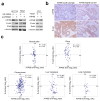
ATP synthase epsilon subunit (ATP5E) expression induces the epithelial-to-mesenchymal transition. (a) Western blot analysis of E-cadherin and Snail expressions upon ATP5E knockdown and overexpression. (b) IHC staining of ATP5E and E-cadherin in serial sections of colon cancer specimens. (c) ATP5E and E-cadherin expression profiles of normal colon, polyp, primary colon tumor, liver metastatic tumor, and lung metastatic tumor tissues in the GSE41258 microarray dataset.
Figure 4
Figure 4
ATP synthase epsilon subunit (ATP5E) upregulation connects the adenosine monophosphate-activated protein kinase (AMPK)-AKT-hypoxia-inducible factor-1α (HIF1a) signaling axis to the epithelial-to-mesenchymal transition (EMT) phenotype in lung metastatic tumors. (a) Upstream regulator analysis of differentially expressed genes in lung metastatic tumors from the GSE41258 dataset. The orange circle indicates predicted activation, while the blue circle indicates predicted inhibition. (b) Differentially expressed genes downstream of AKT and HIF1a extracted from the GSE41258 dataset. (c) Western blot analysis of the phosphorylation status of AMPK and AKT upon ATP5E inhibition in HCT116 and H3347 cells. (d) Western blot analysis of the HIF1a protein upon ATP5E inhibition in hypoxia.

References

    1. Ferlay J., Soerjomataram I., Dikshit R., Eser S., Mathers C., Rebelo M., Parkin D.M., Forman D., Bray F. Cancer incidence and mortality worldwide: Sources, methods and major patterns in GLOBOCAN 2012. Int. J. Cancer. 2015;136:E359–E386. doi: 10.1002/ijc.29210. - DOI - PubMed
    1. Jemal A., Bray F., Center M.M., Ferlay J., Ward E., Forman D. Global cancer statistics. CA Cancer J. Clin. 2011;61:69–90. doi: 10.3322/caac.20107. - DOI - PubMed
    1. Griffin M.R., Bergstralh E.J., Coffey R.J., Beart R.W., Jr., Melton L.J., 3rd Predictors of survival after curative resection of carcinoma of the colon and rectum. Cancer. 1987;60:2318–2324. doi: 10.1002/1097-0142(19871101)60:9<2318::AID-CNCR2820600934>3.0.CO;2-B. - DOI - PubMed
    1. Sargent D.J., Patiyil S., Yothers G., Haller D.G., Gray R., Benedetti J., Buyse M., Labianca R., Seitz J.F., O’Callaghan C.J., et al. End Points for Colon Cancer Adjuvant Trials: Observations and Recommendations Based on Individual Patient Data from 20,898 Patients Enrolled Onto 18 Randomized Trials from the ACCENT Group. J. Clin. Oncol. 2007;25:4569–4574. doi: 10.1200/JCO.2006.10.4323. - DOI - PubMed
    1. Bates R.C., Mercurio A.M. The epithelial-mesenchymal transition (EMT) and colorectal cancer progression. Cancer Biol. Ther. 2005;4:365–370. - PubMed