

The Intrinsic GDP/GTP Exchange Activities of Cdc42 and Rac1 Are Critical Determinants for Their Specific Effects on Mobilization of the Actin Filament System
- PMID: 31330900
- PMCID: PMC6678527
- DOI: 10.3390/cells8070759
The Intrinsic GDP/GTP Exchange Activities of Cdc42 and Rac1 Are Critical Determinants for Their Specific Effects on Mobilization of the Actin Filament System
Abstract
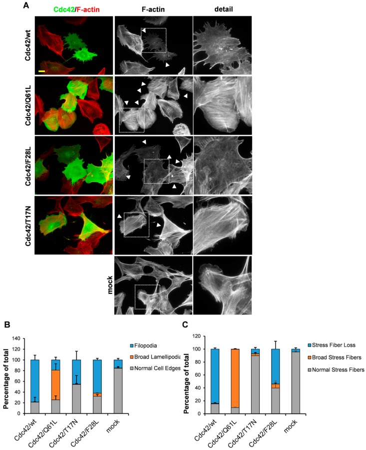
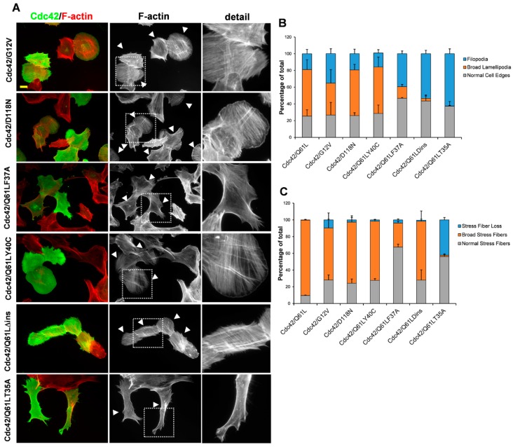
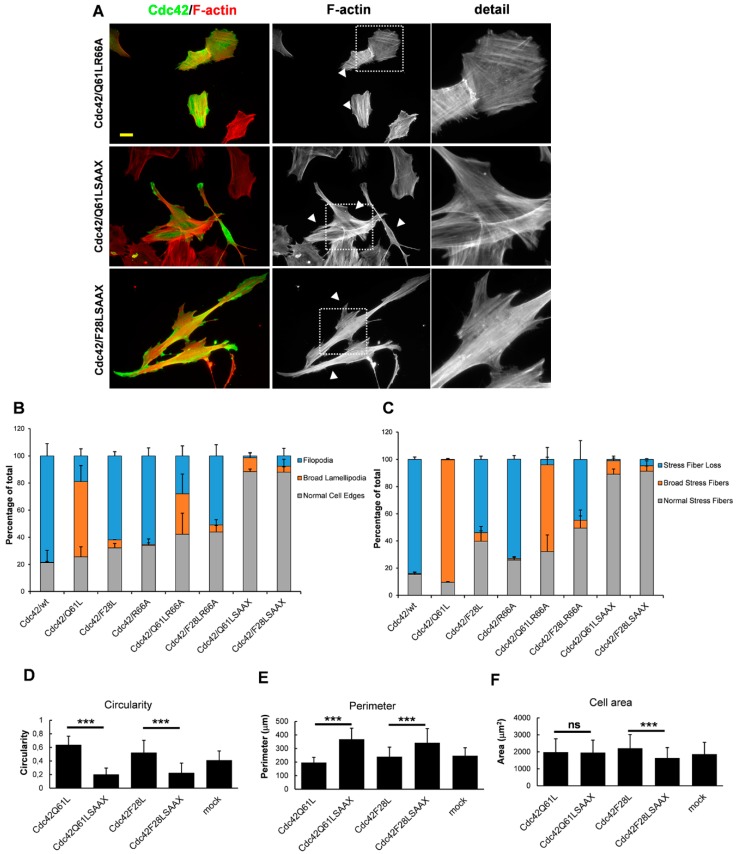
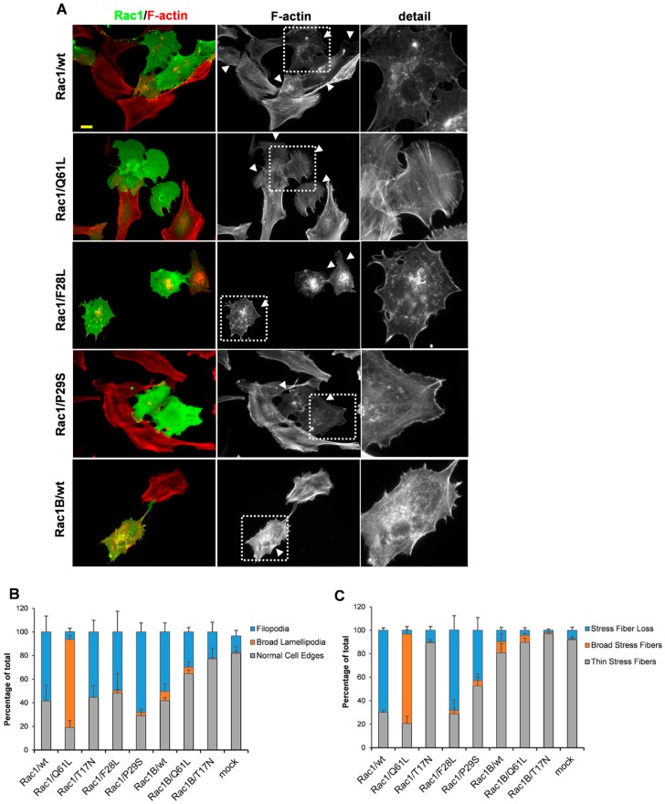
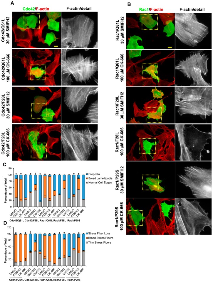
The Rho GTPases comprise a subfamily of the Ras superfamily of small GTPases. Their importance in regulation of cell morphology and cell migration is well characterized. According to the prevailing paradigm, Cdc42 regulates the formation of filopodia, Rac1 regulates the formation of lamellipodia, and RhoA triggers the assembly of focal adhesions. However, this scheme is clearly an oversimplification, as the Rho subfamily encompasses 20 members with diverse effects on a number of vital cellular processes, including cytoskeletal dynamics and cell proliferation, migration, and invasion. This article highlights the importance of the catalytic activities of the classical Rho GTPases Cdc42 and Rac1, in terms of their specific effects on the dynamic reorganization of the actin filament system. GTPase-deficient mutants of Cdc42 and Rac1 trigger the formation of broad lamellipodia and stress fibers, and fast-cycling mutations trigger filopodia formation and stress fiber dissolution. The filopodia response requires the involvement of the formin family of actin nucleation promotors. In contrast, the formation of broad lamellipodia induced by GTPase-deficient Cdc42 and Rac1 is mediated through Arp2/3-dependent actin nucleation.
Keywords: Cdc42; Rac; Rho GTPases; actin; fast-cycling; filopodia.
Conflict of interest statement
The author declares no conflict of interest.
Figures
Similar articles
-
DEF6, a novel PH-DH-like domain protein, is an upstream activator of the Rho GTPases Rac1, Cdc42, and RhoA.Exp Cell Res. 2004 Apr 1;294(2):335-44. doi: 10.1016/j.yexcr.2003.12.004. Exp Cell Res. 2004. PMID: 15023524
-
Cross-talk between Rac1 and Cdc42 GTPases regulates formation of filopodia required for dengue virus type-2 entry into HMEC-1 cells.J Gen Virol. 2009 Dec;90(Pt 12):2902-2911. doi: 10.1099/vir.0.014159-0. Epub 2009 Aug 26. J Gen Virol. 2009. PMID: 19710257
-
Coactivation of Rac1 and Cdc42 at lamellipodia and membrane ruffles induced by epidermal growth factor.Mol Biol Cell. 2004 Mar;15(3):1003-10. doi: 10.1091/mbc.e03-08-0609. Epub 2003 Dec 29. Mol Biol Cell. 2004. PMID: 14699061 Free PMC article.
-
Regulation of phosphorylation pathways by p21 GTPases. The p21 Ras-related Rho subfamily and its role in phosphorylation signalling pathways.Eur J Biochem. 1996 Dec 1;242(2):171-85. doi: 10.1111/j.1432-1033.1996.0171r.x. Eur J Biochem. 1996. PMID: 8973630 Review.
-
Inhibition of Cdc42 and Rac1 activities in pheochromocytoma, the adrenal medulla tumor.Small GTPases. 2017 Apr 3;8(2):122-127. doi: 10.1080/21541248.2016.1202634. Epub 2016 Jun 29. Small GTPases. 2017. PMID: 27355516 Free PMC article. Review.
Cited by
-
AKBA Promotes Axonal Regeneration via RhoA/Rictor to Repair Damaged Sciatic Nerve.Int J Mol Sci. 2022 Dec 14;23(24):15903. doi: 10.3390/ijms232415903. Int J Mol Sci. 2022. PMID: 36555556 Free PMC article.
-
A deep generative model for deciphering cellular dynamics and in silico drug discovery in complex diseases.Nat Biomed Eng. 2025 Jun 20. doi: 10.1038/s41551-025-01423-7. Online ahead of print. Nat Biomed Eng. 2025. PMID: 40542107
-
Fam20c regulates the calpain proteolysis system through phosphorylating Calpasatatin to maintain cell homeostasis.J Transl Med. 2023 Jun 27;21(1):417. doi: 10.1186/s12967-023-04275-4. J Transl Med. 2023. PMID: 37370126 Free PMC article.
-
Rabenosyn-5 suppresses non-small cell lung cancer metastasis via inhibiting CDC42 activity.Cancer Gene Ther. 2024 Oct;31(10):1465-1476. doi: 10.1038/s41417-024-00813-4. Epub 2024 Jul 29. Cancer Gene Ther. 2024. PMID: 39075137 Free PMC article.
-
Spatiotemporal Coordination of Rac1 and Cdc42 at the Whole Cell Level during Cell Ruffling.Cells. 2023 Jun 15;12(12):1638. doi: 10.3390/cells12121638. Cells. 2023. PMID: 37371108 Free PMC article.
References
Publication types
MeSH terms
Substances
LinkOut - more resources
Full Text Sources
Research Materials
Miscellaneous
