. 2019 Jul 19;9(7):170.
doi: 10.3390/brainsci9070170.

White Matter Hyperintensity Regression: Comparison of Brain Atrophy and Cognitive Profiles with Progression and Stable Groups

Affiliations

White Matter Hyperintensity Regression: Comparison of Brain Atrophy and Cognitive Profiles with Progression and Stable Groups

Omar M Al-Janabi et al. Brain Sci. .

Abstract

Subcortical white matter hyperintensities (WMHs) in the aging population frequently represent vascular injury that may lead to cognitive impairment. WMH progression is well described, but the factors underlying WMH regression remain poorly understood. A sample of 351 participants from the Alzheimer's Disease Neuroimaging Initiative 2 (ADNI2) was explored who had WMH volumetric quantification, structural brain measures, and cognitive measures (memory and executive function) at baseline and after approximately 2 years. Selected participants were categorized into three groups based on WMH change over time, including those that demonstrated regression (n = 96; 25.5%), stability (n = 72; 19.1%), and progression (n = 209; 55.4%). There were no significant differences in age, education, sex, or cognitive status between groups. Analysis of variance demonstrated significant differences in atrophy between the progression and both regression (p = 0.004) and stable groups (p = 0.012). Memory assessments improved over time in the regression and stable groups but declined in the progression group (p = 0.003; p = 0.018). WMH regression is associated with decreased brain atrophy and improvement in memory performance over two years compared to those with WMH progression, in whom memory and brain atrophy worsened. These data suggest that WMHs are dynamic and associated with changes in atrophy and cognition.

Keywords: ADNI, brain atrophy, cognition; Stable WMH; WMH progression; WMH regression; white matter hyperintensities.


Conflict of interest statement

The authors declare no conflict of interest.

Figures
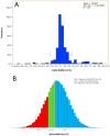
Figure 1
Figure 1
White matter hyperintensity distribution in the sample studied. (A) The true distribution of the data, showing notable leptokurtosis. Black arrows indicate standard deviation, demonstrating why standard deviation was not deemed an appropriate criterion for separating groups. (B) Divisions of the three white matter hyperintensity (WMH) groups. Visualization only.
Figure 2
Figure 2
Descriptive figure showing the dynamic nature of the white matter hyperintensity changes over time and the associated change in brain volume and cognitive performance. The coronal image cartoon represents the brain and ventricle size (scale of change exaggerated for emphasis), while the battery represents memory performance (improved memory corresponding to more battery capacity and green). The first column represents a normal brain with no WMH and normal memory, the second and third columns the baseline and two-year follow up showing changes over the course of the study. The upper row represents WMH stability. Brain volume and memory remained stable in the follow up visit as shown in A2 and A3. The middle row illustrates the case of progression of WMH over time. In B3, the WMH region enlarges, brain volume decreases along with ventricular increases and memory declines compared to B2. Finally, the lower row represents cases of WMH regression over time. In C3, the WMH lesion volume shrinks, brain volume is maintained, and memory is improved when compared to C2.
Figure 3
Figure 3
Conceptual descriptive figure showing the possible mechanisms that lead to white matter hyperintensity regression overtime and their associated effect on the brain volume and the cognitive performance (same conventions as in Figure 2). The upper row demonstrates the first potential mechanism of white matter hyperintensity (WMH) regression due to ischemic non-reversible injury. In A3, the WMH lesion contracts (black arrows) resulting in brain volume reduction compared to A2. Because the contraction is merely mechanical, memory performance is not changed compared to A2. The middle row illustrates inflammation as a potential cause of WMH regression. In B3, the edema resolves, the brain volume consequently decreases but memory is improved compared to B2. The difference between A3 and B3 is in expected memory performance. Lower row represents a third explanation of WMH regression, most consistent with our data. In C3, WMH lesion volume regresses, but brain volume and memory performance increase compared to C2. The difference between C3 and B3 is in brain atrophy, which is greater in B3.

References

    1. Pantoni L., Garcia J.H. The significance of cerebral white matter abnormalities 100 years after binswanger’s report: A review. Stroke. 1995;26:1293–1301. doi: 10.1161/01.STR.26.7.1293. - DOI - PubMed
    1. Schmidt R., Fazekas F., Kapeller P., Schmidt H., Hartung H.-P. Mri white matter hyperintensities three-year follow-up of the austrian stroke prevention study. Neurology. 1999;53:132. doi: 10.1212/WNL.53.1.132. - DOI - PubMed
    1. Veldink J.H., Scheltens P., Jonker C., Launer L.J. Progression of cerebral white matter hyperintensities on mri is related to diastolic blood pressure. Neurology. 1998;51:319–320. doi: 10.1212/WNL.51.1.319. - DOI - PubMed
    1. Arvanitakis Z., Fleischman D.A., Arfanakis K., Leurgans S.E., Barnes L.L., Bennett D.A. Association of white matter hyperintensities and gray matter volume with cognition in older individuals without cognitive impairment. Brain Struct. Funct. 2016;221:2135–2146. doi: 10.1007/s00429-015-1034-7. - DOI - PMC - PubMed
    1. Van den Berg E., Geerlings M.I., Biessels G.J., Nederkoorn P.J., Kloppenborg R.P. White matter hyperintensities and cognition in mild cognitive impairment and alzheimer’s disease: A domain-specific meta-analysis. J. Alzheimers Dis. 2018;63:515–527. doi: 10.3233/JAD-170573. - DOI - PubMed

LinkOut - more resources