Review
. 2019 Jul 19;8(7):747.
doi: 10.3390/cells8070747.

Macrophage-Mediated Subversion of Anti-Tumour Immunity

Affiliations
Review

Macrophage-Mediated Subversion of Anti-Tumour Immunity

Valeria Quaranta et al. Cells. .

Abstract

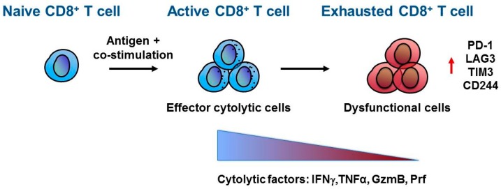
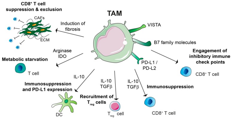
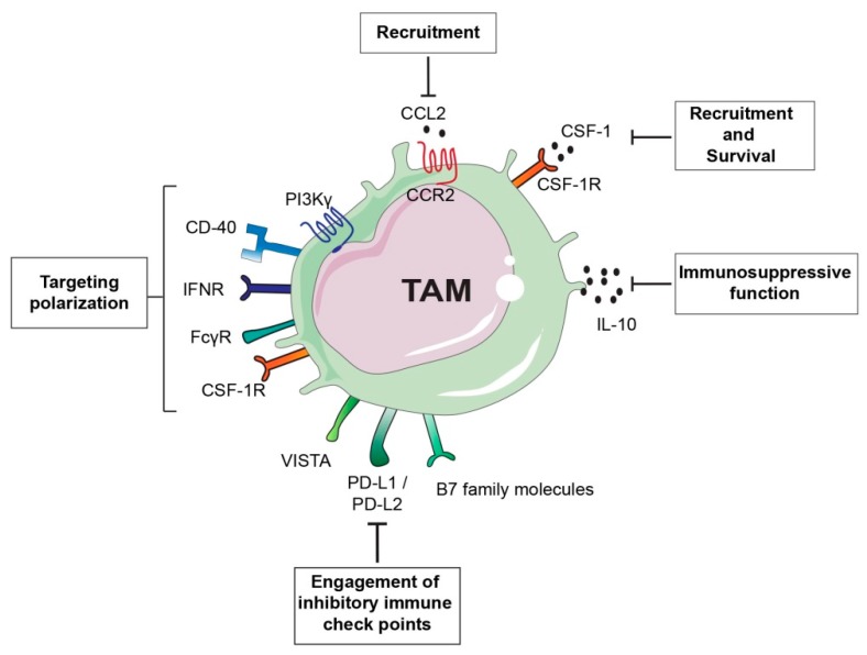
Despite the incredible clinical benefits obtained by the use of immune checkpoint blockers (ICBs), resistance is still common for many types of cancer. Central for ICBs to work is activation and infiltration of cytotoxic CD8+ T cells following tumour-antigen recognition. However, it is now accepted that even in the case of immunogenic tumours, the effector functions of CD8+ T cells are highly compromised by the presence of an immunosuppressive tumour microenvironment (TME) at the tumour site. Tumour-associated macrophages (TAMs) are among the most abundant non-malignant stromal cell types within the TME and they are crucial drivers of tumour progression, metastasis and resistance to therapy. TAMs are able to regulate either directly or indirectly various aspects of tumour immunity, including T cell recruitment and functions. In this review we discuss the mechanisms by which TAMs subvert CD8+ T cell immune surveillance and how their targeting in combination with ICBs represents a very powerful therapeutic strategy.

Keywords: anti-tumour immunity; cancer; immune checkpoint blockers; immunosuppression; macrophage.


Conflict of interest statement

The authors declare no conflict of interest.

Figures

Figure 1
Figure 1
CD8+ T cell exhaustion. Naïve CD8+ T cells differentiate and activate into effector CD8+ T cells upon antigen recognition and co-stimulation mediated by antigen presenting cells (APC). Once at the tumour site, active CD8+ T cells recognize cancer cells by T cell receptor (TCR)–tumour antigen interaction and direct their effector anti-tumour activity through secretion of cytokines and lytic molecules such as interferon γ (IFNγ), tumor necrosis factor α (TNFα), granzyme B (GzmB) and perforin (Prf). However, within the tumour microenvironment, CD8+ T cells can acquire a dysfunctional or exhausted phenotype characterized by a progressive loss in cytolytic factors and increased expression of inhibitory receptors, including Programmed cell death-1 (PD-1), lymphocyte-activation gene 3 (LAG3), T-cell immunoglobulin and mucin-domain containing 3 (TIM-3), and CD244.
Figure 2
Figure 2
Immunosuppressive role of tumour-associated macrophages (TAMs). TAMs promote immunosuppression by different mechanisms: TAMs can express programmed cell death ligand 1 and ligand 2 (PD-L1 and PD-L2), which bind the T cell inhibitory receptor programmed cell death protein 1 (PD-1), thereby inducing T cell exhaustion. TAM expression of V-domain Ig suppressor of T cell activation (VISTA) and B7 family molecules, such as B7-H4, can have similar function. TAMs secrete immunosuppressive cytokines such as IL-10 and TGFβ, which can have direct suppressive functions on CD8+ T cells, or they can induce recruitment of immunosuppressive CD4+ T regulatory (Treg) cells. TAM secreted IL-10 can impair T cells effector functions indirectly either by inhibit dendritic cell (DC) anti-tumorigenic role or by inducing DC expression of PD-L1. Moreover, amino acid metabolism in TAM results in production of arginase and immunosuppressive metabolites via the indoleamine 2,3-dioxygenase (IDO) pathway, which is responsible of metabolic starvation in T cells. TAMs can also indirectly induce immunosuppression by promoting fibrosis. Indeed cancer associated fibroblast (CAF) secreted factors and/or extracellular matrix (ECM) stiffness are responsible for CD8+ T cell immunosuppression and/or exclusion from the tumour nest.
Figure 3
Figure 3
Tumour-associated macrophage (TAM)-targeted therapeutic approaches. TAM-centered approaches either aim at inhibiting TAM recruitment and survival or they focus on promoting TAM anti-tumorigenic activity. For example inhibition of the monocyte chemoattractant chemokine (C-C motif) ligand 2 (CCL2) or its receptor, as well as blockade of colony stimulation factor (CSF-1)/CSF-1 receptor (CSF-1R) axis can prevent TAM accumulation at the tumour site or TAM survival. CSF-1/CSF-1R blockade can also have the potential to switch TAM phenotype from a pro-tumorigenic M2-like to a pro-inflammatory M1-like. Similarly, approaches aimed at re-educating TAMs toward an anti-tumorigenic phenotype are successful as in the case of the use of inhibitors of gamma isoform of phosphoinositide 3-kinase (PI3Kγ) or Ig receptor gamma (FcRγ). Interferon gamma (IFNγ) administration or activation of the stimulatory receptor CD40 using anti-CD40 antibody agonists can induce re-education of pro-tumorigenic TAMs. Another alternative approach is the targeting the immunosuppressive function of TAMs by inhibiting IL-10 or blockade of immune checkpoint receptors engagement.

Similar articles

Cited by

References

    1. Hanahan D., Weinberg R.A. Hallmarks of cancer: The next generation. Cell. 2011;144:646–674. doi: 10.1016/j.cell.2011.02.013. - DOI - PubMed
    1. Joyce J.A., Pollard J.W. Microenvironmental regulation of metastasis. Nat. Rev. Cancer. 2009;9:239–252. doi: 10.1038/nrc2618. - DOI - PMC - PubMed
    1. Ribas A., Wolchok J.D. Cancer immunotherapy using checkpoint blockade. Science (80-). 2018;359:1350–1355. doi: 10.1126/science.aar4060. - DOI - PMC - PubMed
    1. Chen D.S., Mellman I. Oncology meets immunology: The cancer-immunity cycle. Immunity. 2013;39:1–10. doi: 10.1016/j.immuni.2013.07.012. - DOI - PubMed
    1. Hanahan D., Coussens L.M.M. Accessories to the crime: Functions of cells recruited to the tumor microenvironment. Cancer Cell. 2012;21:309–322. doi: 10.1016/j.ccr.2012.02.022. - DOI - PubMed

Publication types

Substances