Review
. 2019 Oct;98(11):1186-1194.
doi: 10.1177/0022034519864592. Epub 2019 Jul 22.

Epithelial Cell Lineage and Signaling in Murine Salivary Glands

Affiliations
Review

Epithelial Cell Lineage and Signaling in Murine Salivary Glands

M H Aure et al. J Dent Res. 2019 Oct.

Abstract

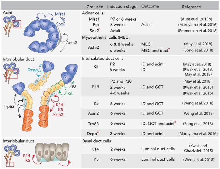
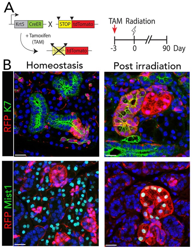
Maintaining salivary gland function is critical for oral health. Loss of saliva is a common side effect of therapeutic irradiation for head and neck cancer or autoimmune diseases such as Sjögren's syndrome. There is no curative treatment, and current strategies proposed for functional regeneration include gene therapy to reengineer surviving salivary gland tissue, cell-based transplant therapy, use of bioengineered glands, and development of drugs/biologics to stimulate in vivo regeneration or increase secretion. Understanding the genetic and cellular mechanisms required for development and homeostasis of adult glands is essential to the success of these proposed treatments. Recent advances in genetic lineage tracing provide insight into epithelial lineage relationships during murine salivary gland development. During early fetal gland development, epithelial cells expressing keratin 14 (K14) Sox2, Sox9, Sox10, and Trp63 give rise to all adult epithelium, but as development proceeds, lineage restriction occurs, resulting in separate lineages of myoepithelial, ductal, and acinar cells in postnatal glands. Several niche signals have been identified that regulate epithelial development and lineage restriction. Fibroblast growth factor signaling is essential for gland development, and other important factors that influence epithelial patterning and maturation include the Wnt, Hedgehog, retinoic acid, and Hippo signaling pathways. In addition, other cell types in the local microenvironment, such as endothelial and neuronal cells, can influence epithelial development. Emerging evidence also suggests that specific epithelial cells will respond to different types of salivary gland damage, depending on the cause and severity of damage and the resulting damaged microenvironment. Understanding how regeneration occurs and which cell types are affected, as well as which signaling factors drive cell lineage decisions, provides specific targets to manipulate cell fate and improve regeneration. Taken together, these recent advances in understanding cell lineages and the signaling factors that drive cell fate changes provide a guide to develop novel regenerative treatments.

Keywords: epithelial signaling; lineage tracing; progenitor; regeneration; salivary gland development; submandibular gland.


Conflict of interest statement

The authors declare no potential conflicts of interest with respect to the authorship and/or publication of this article.

Figures

Figure 1.
Figure 1.
Using genetic models to investigate lineage relationships. Lineage tracing is dependent on knowing the temporospatial expression pattern of the gene of interest driving Cre expression. Expression of Gene-X has a dynamic stage-dependent localization pattern. Outcomes of lineage tracing of Gene-X are dependent on the experimental design. Inducing Gene-X-Cre at stage 1 (green) will lineage trace every cell at stage 3, while induction at stage 2 will lineage trace a subpopulation at stage 3. Both outcomes differ from the expression pattern of Gene-X at stage 3.
Figure 2.
Figure 2.
Genetic labeling of AQP5 lineage. (A) Crossing the ACID (Aqp5-Cre-IRES-DsRed) mouse line with the mTmG reporter line leads to constitutive expression of Cre in AQP5+ cells and their progeny. Cre induction mediates recombination by excising the membrane-targeted Tomato (mT) sequence, leading to expression of membrane-targeted GFP (mG). (B) AQP5-Cre is expressed in acinar cells, leading to mG (green) expression in these cells. (C) Cells that are AQP5 negative express mT (red), which includes myoepithelial and duct cells. (D) The overlay shows mG expression in acinar cells (green) and mT (red) expression in other lineages. Arrow indicates intercalated duct; arrowhead indicates myoepithelial cell. Scale bar = 20 µm.
Figure 3.
Figure 3.
Lineage-tracing embryonic glands. Using Cre mouse lines that label the salivary gland anlagen from embryonic day 9.5 (E9.5) to E10.5 lineage traces to the entire gland, confirming that these cells give rise to the entire gland. Inducing lineage tracing from E11.5 to E12.5 gives different results depending on the cells that are traced. MEC, myoepithelial cell; SLG, sublingual gland; SMG, submandibular gland.
Figure 4.
Figure 4.
Sox10 is required for secretory cell fate. (A, B) Genetic ablation of Sox10 in epithelial cells leads to loss of Kit+ progenitors (arrow) in embryonic day 16 endbuds. Loss of Sox10 also led to a decrease in proliferation (CCND1) in the epithelium (CDH1). (C, D) Comparing control glands with Sox10fl/fl shows loss of differentiating acinar cells (identified by expression of SMGc and AQP5). AQP5, aquaporin 5; CCND1, cyclin D1; CDH1, cadherin 1; KIT, KIT proto-oncogene receptor tyrosine kinase; SMGc, submandibular gland protein C. Scale bar: 20 µm. From Figure 4 of Athwal et al. (2019).
Figure 5.
Figure 5.
Lineage tracing in adult glands. Duct, myoepithelial, and acinar cells are mainly maintained by separate lineages. 1Sox2 lineage tracing in adult mice is specific to sublingual gland. 2A modest number of duct cells were lineage traced from Acta2+ cells in this study. 3A modest number of acinar cells were lineage traced from Trp63 following long-term chase in the submandibular gland (SMG). 4In parotid gland (PG) specifically, Dcpp+ intercalated ducts (IDs) infrequently also give rise to acinar cells. Dcpp, demilune cell and parotid protein 1; GCT, granular convoluted tubules; P, postnatal day. Dotted arrows indicate age-specific or rare events reported.
Figure 6.
Figure 6.
Lineage plasticity after irradiation. (A) Tamoxifen (TAM)–inducible K5CreER mice were crossed with the R26tdTomato reporter strain to follow K5 lineage in postnatal glands. Tamoxifen treatment induces Cre-mediated recombination in K5+ cells, which excises the stop codon allowing tdTomato (RFP) expression in the K5+ cells and their descendants. Experimental timeline for tamoxifen-dependent Cre induction, radiation, and tissue harvest. Mice were irradiated 3 d after tamoxifen treatment, and irradiated glands were compared with nonirradiated glands following a 90-d chase period. (B) After 90 d, the expanded number of RFP-labeled cells in female submandibular glands remain colocalized with K7, a specific duct marker, but not with Mist1, an acinar cell marker. However, 90 d after irradiation, RFP labels clusters of cells that colocalize with duct and acinar cells, indicating that K5 cells have given rise to duct and acini following irradiation. Nuclei are stained with DAPI (blue). Scale bars: 25 µm. From Figures 1 and 4 of Weng et al. (2018).

References

    1. Abashev TM, Metzler MA, Wright DM, Sandell LL. 2017. Retinoic acid signaling regulates Krt5 and Krt14 independently of stem cell markers in submandibular salivary gland epithelium. Dev Dyn. 246(2):135–147. - PMC - PubMed
    1. Athwal HK, Murphy G, Tibbs E, Cornett A, Hill E, Yeoh K, Berenstein E, Hoffman MP, Lombaert IMA. 2019. SOX10 regulates plasticity of epithelial progenitors toward secretory units of exocrine glands. Stem Cell Rep. 12(2):366–380. - PMC - PubMed
    1. Aure MH, Arany S, Ovitt CE. 2015. Salivary glands: stem cells, self-duplication, or both? J Dent Res. 94(11):1502–1507. - PMC - PubMed
    1. Aure MH, Konieczny SF, Ovitt CE. 2015. Salivary gland homeostasis is maintained through acinar cell self-duplication. Dev Cell. 33(2):231–237. - PMC - PubMed
    1. Bullard T, Koek L, Roztocil E, Kingsley PD, Mirels L, Ovitt CE. 2008. Ascl3 expression marks a progenitor population of both acinar and ductal cells in mouse salivary glands. Dev Biol. 320(1):72–78. - PMC - PubMed

Publication types