. 2019 Jul 22;20(1):597.
doi: 10.1186/s12864-019-5969-6.

Cold seawater induces early sexual developmental stages in the BPG axis of European eel males

Affiliations

Cold seawater induces early sexual developmental stages in the BPG axis of European eel males

Christoffer Rozenfeld et al. BMC Genomics. .

Abstract

Background: The impossibility of closing the life cycle of the European eel (Anguilla anguilla) in captivity troubles the future of this critically endangered species. In addition, the European eel is a highly valued and demanded resource, thus the successful closing of its life cycle would have a substantial economic and ecological impact. With the aim of obtaining the highest gamete quality, the study of the effects of environmental factors, such as temperature, on reproductive performance may prove valuable. This is especially true for the exposure to cold water, which has been reported to improve sexual development in multiple other Actinopterygii species.

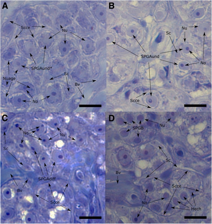
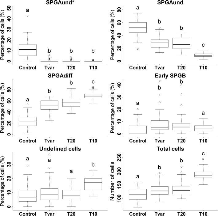
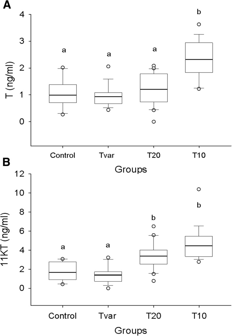
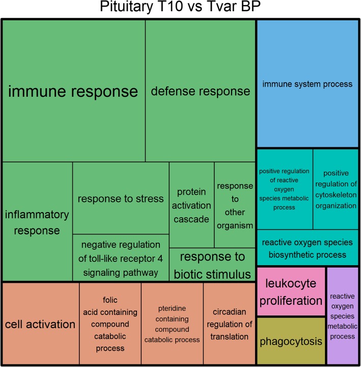
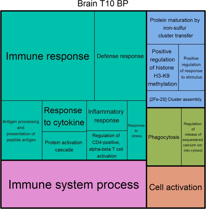
Results: European eel males treated with cold seawater (10 °C, T10) for 2 weeks showed an increase in the proliferation and differentiation of spermatogonial cells until the differentiated spermatogonial type A cell stage, and elevated testosterone and 11-ketotestosterone plasma levels. Transcriptomes from the tissues of the brain-pituitary-gonad (BPG) axis of T10 samples revealed a differential gene expression profile compared to the other experimental groups, with clustering in a principal component analysis and in heat maps of all differentially expressed genes. Furthermore, a functional analysis of differentially expressed genes revealed enriched gene ontology terms involved in the regulation of circadian rhythm, histone modification, meiotic nuclear division, and others.

Conclusions: Cold seawater treatment had a clear effect on the activity of the BPG-axis of European eel males. In particular, our cold seawater treatment induces the synchronization and increased proliferation and differentiation of specific spermatogonial cells. In the transcriptomic results, genes related to thermoception were observed. This thermoception may have caused the observed effects through epigenetic mechanisms, since all analysed tissues further revealed differentially expressed genes involved in histone modification. The presented results support our hypothesis that a low temperature seawater treatment induces an early sexual developmental stage in European eels. This hypothesis is logical given that the average temperature experienced by eels in the early stages of their oceanic reproductive migration is highly similar to that of this cold seawater treatment. Further studies are needed to test whether a cold seawater treatment can improve the response of European eels to artificial hormonal treatment, as the results suggest.

Keywords: Anguilla anguilla; Epigenetics; Histology; Immunofluorescence; Migration; RNA-sequencing; Radioimmunoassay; Spermatogonial proliferation; Temperature.


Conflict of interest statement

The authors declare that they have no competing interests.

Figures

Fig. 1
Fig. 1
Identified spermatogonia types. Forty times magnification fields of selected histological sections representing the spermatogonia stages: the most undifferentiated spermatogonia type A (SPGAund*; panel a), the second most undifferentiated spermatogonia type A (SPGAund; panel b), differentiated spermatogonia type A cells (SPGAdiff; panel c), and early spermatogonia type B cells (SPGB; panel d). These identifiable characteristics are further labelled with arrows: Blood vessels (Bv), nucleus (Nu), nucleoli (No), Sertoli cells (Sc), Sertoli cell cytoplasmic extensions (Scce), heterochromatin (Hech) and nuage (Nuage)
Fig. 2
Fig. 2
Histological cell counts. Boxplots of cell counting results for the T10, T20, Tvar and Control groups. The panels show the percentage proportion of the most undifferentiated spermatogonia type A cells (SPGAund*), the second most undifferentiated spermatogonia type A cells (SPGAund), the differentiated spermatogonia type A cells (SPGAdiff), and the early spermatogonia type B cells (Early SPGB) in each group. The panels labelled “Undefined cells” presents the percentage proportion of cells in each group, which were identified as spermatogonial cells but could not be distinguished between the specific spermatogonial cell types. The panels labelled “Total cells” presents the accumulated cells count of all identified cell types in each group. Letters indicate significant differences
Fig. 3
Fig. 3
Radioimmunoassay steroid results. Boxplots of radioimmunoassay steroid results from the blood of fish from the T10, T20, Tvar and control groups. Significant differences are indicated with letters. Panel a shows the testosterone (T) results, while panel b shows the 11-ketotestosterone (11KT) results
Fig. 4
Fig. 4
Pituitary Lhβ histochemical identification. Confocal images of the immunofluorescence labelled European eel (Anguilla anguilla) male pituitaries, which showed the strongest Lhβ signal from each of the analyzed groups: the 2 week 10 °C pretreated group (T10; panel d, e, and f), the 2 week 20 °C pretreated group (T20; panel g, h, and i), and Control (Panel a, b, and c). “DAPI” indicates pictures filtered to only reveal fluorescents labeled to 4,6-diamidino-2-phenylindole dihydrochloride (Panel a, d, and g). “Lhβ” indicates pictures filtered to only reveal fluorescents labeled to luteinizing hormone beta subunit protein (Panel b, e, and h). “DAPI / Lhβ” indicates pictures filtered to reveal both fluorescents labelled to luteinizing hormone beta subunit protein and 4,6-diamidino-2-phenylindole dihydrochloride (Panel c, f, and i)
Fig. 5
Fig. 5
Principal component analysis of expression data. Principal component 2 (PC2) over principal component 1 (PC1) from 3 principal component analysis of all normalized expression data from all transcriptomes of the pituitary, testes, and brain samples. Hollow circles labeled “Cont.”, “T20”, and “Tvar” represents transcriptomes of Control, T20 and Tvar samples, respectively. T10 samples are marked with red filled circles, and labelled “T10”
Fig. 6
Fig. 6
Treemap of BP GO terms found between T10 and Tvar in the pituitary. Treemap of the significantly enriched biological process GO term from the significantly differentially expressed genes found between the T10 and Tvar groups from the pituitary samples. Each rectangle represents a single cluster of related terms. Loosely related single cluster rectangles are clustered together in superclusters of the same color. The size of each cluster is adjusted to reflect the false discovery rate corrected P-value (FDR) of the enrichment of the GO tem (larger rectangles indicates lower FDR)
Fig. 7
Fig. 7
Treemap of BP GO terms found between T10 and Control in the brain. Treemap of the significantly enriched biological process GO terms from the significantly differentially expressed genes found between the T10 and Control groups from the brain samples. Each rectangle represents a single cluster of related terms. Loosely related single cluster rectangles are clustered together in superclusters of the same color. The size of each cluster is adjusted to reflect the false discovery rate corrected P-value (FDR) of the enrichment of the GO terms (larger rectangles indicates lower FDR)

References

    1. Jacoby D, Gollock M. Anguilla anguilla. The IUCN Red List of Threatened Species: IUCN; 2014. Version 2014.3. http://www.iucnredlist.org
    1. Peñaranda DS, Gallego V, Rozenfeld C, Herranz-Jusdado JG, Pérez L, Gómez A, et al. Using specific recombinant gonadotropins to induce spermatogenesis and spermiation in the European eel (Anguilla anguilla) Theriogenology. 2018;107:6–20. doi: 10.1016/j.theriogenology.2017.11.002. - DOI - PubMed
    1. Pérez L, Asturiano JF, Tomás A, Zegrari S, Barrera R, Espinós FJ, et al. Induction of maturation and spermiation in the male European eel: assessment of sperm quality throughout treatment. J Fish Biol. 2000;57(6):1488–1504. doi: 10.1111/j.1095-8649.2000.tb02227.x. - DOI
    1. Butts IAE, Sørensen SR, Politis SN, Pitcher TE, Tomkiewicz J. Standardization of fertilization protocols for the European eel, Anguilla anguilla. Aquaculture. 2014;426–427:9–13. doi: 10.1016/j.aquaculture.2014.01.020. - DOI
    1. Mylonas CC, Duncan NJ, Asturiano JF. Hormonal manipulations for the enhancement of sperm production in cultured fish and evaluation of sperm quality. Aquaculture. 2017;472:21–44. doi: 10.1016/j.aquaculture.2016.04.021. - DOI

MeSH terms