. 2019 Jul 22;20(1):164.
doi: 10.1186/s12931-019-1137-4.

The canonical TGF-β/Smad signalling pathway is involved in PD-L1-induced primary resistance to EGFR-TKIs in EGFR-mutant non-small-cell lung cancer

Affiliations

The canonical TGF-β/Smad signalling pathway is involved in PD-L1-induced primary resistance to EGFR-TKIs in EGFR-mutant non-small-cell lung cancer

Yang Zhang et al. Respir Res. .

Abstract

Background: Approximately 30% of patients with epidermal growth factor receptor (EGFR)-activating mutations have no response to EGFR-tyrosine kinase inhibitors (TKIs) (primary resistance). However, little is known about the molecular mechanism involved in primary resistance to EGFR-TKIs in EGFR-mutant non-small cell lung cancer (NSCLC). Programmed death ligand-1 (PD-L1) plays important regulatory roles in intracellular functions and leads to acquired resistance to EGFR-TKIs in NSCLC. Here, we investigated the mechanistic role of PD-L1 in primary resistance to EGFR-TKIs in EGFR-mutant NSCLC cells.

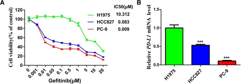
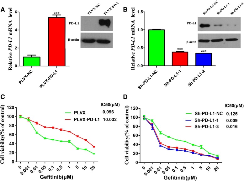
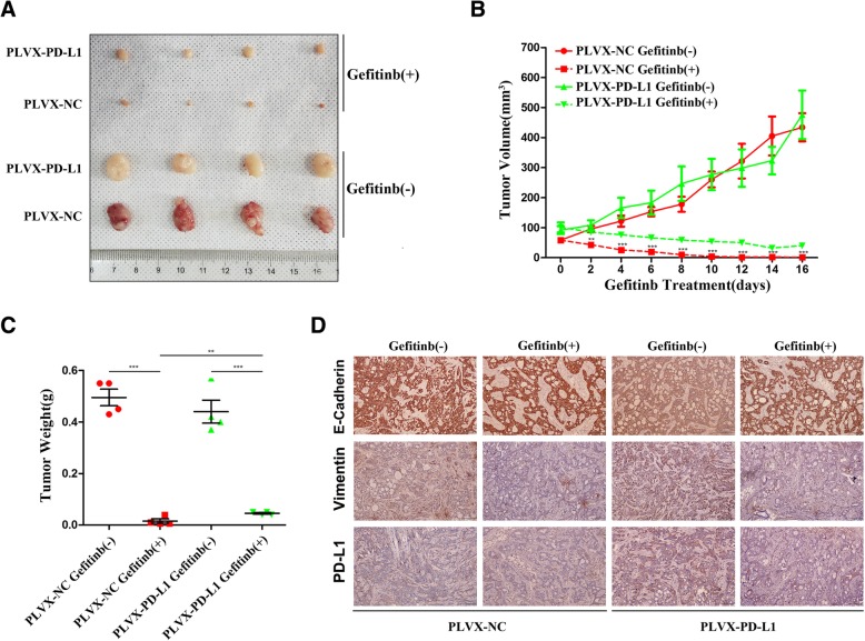
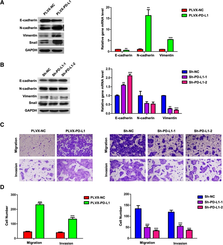
Methods: The expression levels of PD-L1 and the sensitivity to gefitinib in H1975, HCC827 and PC-9 cells were determined by quantitative real-time PCR analysis (qRT-PCR) and Cell Counting Kit-8 (CCK-8) assays, respectively. Molecular manipulations (silencing or overexpression) were performed to assess the effect of PD-L1 on sensitivity to gefitinib, and a mouse xenograft model was used for in vivo confirmation. Western blotting and qRT-PCR were used to analyse the expression of epithelial-mesenchymal transition (EMT) markers. The effect of PD-L1 on migratory and invasive abilities was evaluated using the Transwell assay and mice tail intravenous injection.

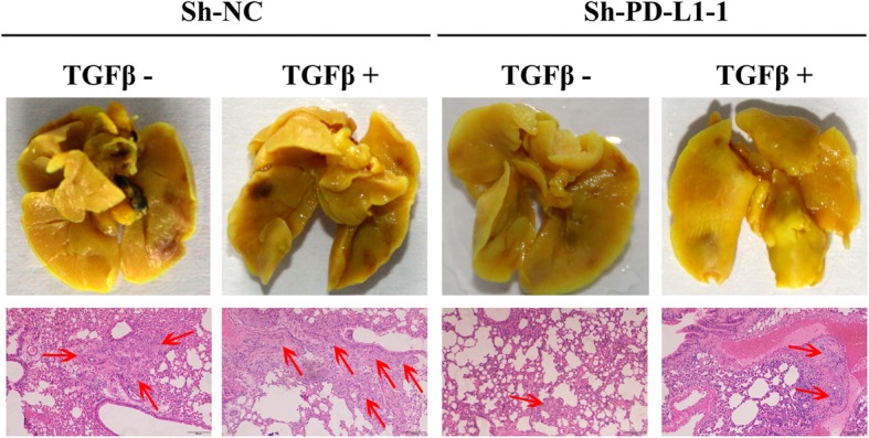
Results: Higher expression of PD-L1 was related to less sensitivity to gefitinib in EGFR-mutant NSCLC cell lines. The overexpression or knockdown of PD-L1 presented diametrical sensitivity to gefitinib in vitro and in vivo. Furthermore, the overexpression of PD-L1 led to primary resistance to gefitinib through the induction of EMT, which was dependent on the upregulation of Smad3 phosphorylation. Moreover, in the mouse model, the knockdown of PD-L1 inhibited transforming growth factor (TGF)-β1-induced cell metastasis in vivo.

Conclusion: PD-L1 contributes to primary resistance to EGFR-TKI in EGFR-mutant NSCLC cells, which may be mediated through the induction of EMT via the activation of the TGF-β/Smad canonical signalling pathway.

Keywords: Drug resistance; EGFR-TKI; NSCLC; PD-L1; TGF-β/Smad signalling.


Conflict of interest statement

The authors declare that they have no competing interests.

Figures

Fig. 1
Fig. 1
Correlation between PD-L1 expression and sensitivity to EGFR-TKIs in EGFR-mutant NSCLC cells (a) Three types of EGFR-mutant NSCLC cells were treated with different concentrations of gefitinib, and the IC50 value was calculated from the dose-response survival curve determined by the CCK-8 assay. b The mRNA expression level of PD-L1 in three types of EGFR-mutant NSCLC cells was detected by qRT-PCR. ***P < 0.001
Fig. 2
Fig. 2
PD-L1 is responsible for generating primary resistance to gefitinib. a PD-L1 mRNA and protein expression levels in stable HCC827 cell lines transfected with a PD-L1-expressing plasmid (PLVX-PD-L1) or a negative control (PLVX-NC). b PD-L1 mRNA and protein expression levels in stable HCC827 cell lines transfected with two PD-L1 ShRNAs (Sh-PD-L1–1 and Sh-PD-L1–2) or a negative control (Sh-PD-L1-NC). c and (d) Stable PD-L1-overexpressing and PD-L1-silenced HCC827 cells were treated with different concentrations of gefitinib, and the IC50 value was calculated from the dose-response survival curve determined by the CCK-8 assay. ***P < 0.001
Fig. 3
Fig. 3
Overexpression of PD-L1 attenuated sensitivity to gefitinib in vivo. a The tumour volume was measured at the indicated time intervals and calculated. See the Methods for details. b and (c) At the end of treatment, the tumours were excised, photographed as indicated and weighed. d Immunohistochemical staining of PD-L1, E-cadherin and vimentin was quantified based on staining intensity. *P < 0.05; **P < 0.01; ***P < 0.001
Fig. 4
Fig. 4
PD-L1 induced EMT and promoted migration and invasion. a and (b) E-cadherin, N- cadherin, vimentin and Snail mRNA and protein expression levels in stable PD-L1-overexpressing or PD-L1-silenced HCC827 cells were detected by qRT-PCR and western blotting, respectively. c and (d) Stable PD-L1-overexpressing or PD-L1-silenced HCC827 cells were allowed to migrate through an 8 μM pore Transwell insert, and the migrated cells were stained and counted in at least three microscopic fields (magnification, 100×). The cells were allowed to invade through Matrigel-coated Transwell membranes, and invasive cells were stained and counted under a light microscope. *P < 0.05; **P < 0.01; ***P < 0.001
Fig. 5
Fig. 5
PD-L1 induced EMT by upregulating Smad3 phosphorylation. a Western blot analysis of p-Smad3 and Smad3 in stable PD-L1-overexpressing or PD-L1-silenced HCC827 cells. The PD-L1 blot is the same blot shown in Fig. 2a and b. b After serum starvation for 24 h, PD-L1-overexpressing and PD-L1-silenced HCC827 cells were treated with SIS3(3 μM) or TGF-β1 (10 ng/ml) for 24 h. The expression of p-Smad3, Smad3, E-cadherin and vimentin was analysed by western blotting. c Knockdown of PD-L1 moderated TGF-β1-induced PAI-1 promoter activation. The relative PAI-1 promoter activities of PD-L1-silenced HCC827 cell lines were measured by a Dual-Luciferase Reporter Assay kit. Relative luciferase activity was expressed as the mean fold change from the basal level ± SD of three independent experiments. d and (e) Stable PD-L1-overexpressing and PD-L1-silenced HCC827 cells were treated with or without SIS3(3 μM) and TGF-β (10 ng/ml) for 24 h, respectively, and allowed to migrate through 8-μM pore Transwell inserts. The migrated cells were stained and counted in at least three microscopic fields (magnification, 100×). The cells were allowed to invade through Matrigel-coated Transwell membranes, and invasive cells were stained and counted under a light microscope
Fig. 6
Fig. 6
Knockdown of PD-L1 inhibits cell migration and invasion in vivo. Surgically resected lung tissues were fixed in Bouin’s fluid, and representative images of micrometastases detected by H&E staining are presented; red arrowheads indicate micrometastases

References

    1. Siegel RL, Miller KD, Jemal A. Cancer statistics, 2018. CA Cancer J Clin. 2018;68:7–30. doi: 10.3322/caac.21442. - DOI - PubMed
    1. Rothschild SI. Targeted therapies in non-small cell lung cancer-beyond EGFR and ALK. Cancers (Basel) 2015;7(2):930–949. doi: 10.3390/cancers7020816. - DOI - PMC - PubMed
    1. Wu YL, Cheng Y, Zhou X, et al. Dacomitinib versus gefitinib as first-line treatment for patients with EGFR-mutation-positive non-small-cell lung cancer (ARCHER 1050): a randomised, open-label, phase 3 trial. Lancet Oncol. 2017;18(11):1454–1466. doi: 10.1016/S1470-2045(17)30608-3. - DOI - PubMed
    1. Yang JJ, Zhou C, Huang Y, et al. Icotinib versus whole-BRAIN irradiation in patients with EGFR-mutant non-small-cell lung cancer and multiple BRAIN metastases (BRAIN): a multicentre, phase 3, open-label, parallel, randomised controlled trial. Lancet Respir Med. 2017;5(9):707–716. doi: 10.1016/S2213-2600(17)30262-X. - DOI - PubMed
    1. Maemondo M, Inoue A, Kobayashi K, et al. Gefitinib or chemotherapy for non-small-cell lung cancer with mutated EGFR. N Engl J Med. 2010;362(25):2380–2388. doi: 10.1056/NEJMoa0909530. - DOI - PubMed

MeSH terms