. 2019 Aug 29;132(16):jcs232124.
doi: 10.1242/jcs.232124.

Dissection of GTPase-activating proteins reveals functional asymmetry in the COPI coat of budding yeast

Affiliations

Dissection of GTPase-activating proteins reveals functional asymmetry in the COPI coat of budding yeast

Eric C Arakel et al. J Cell Sci. .

Abstract

The Arf GTPase controls formation of the COPI vesicle coat. Recent structural models of COPI revealed the positioning of two Arf1 molecules in contrasting molecular environments. Each of these pockets for Arf1 is expected to also accommodate an Arf GTPase-activating protein (ArfGAP). Structural evidence and protein interactions observed between isolated domains indirectly suggest that each niche preferentially recruits one of the two ArfGAPs known to affect COPI, i.e. Gcs1/ArfGAP1 and Glo3/ArfGAP2/3, although only partial structures are available. The functional role of the unique non-catalytic domain of either ArfGAP has not been integrated into the current COPI structural model. Here, we delineate key differences in the consequences of triggering GTP hydrolysis through the activity of one versus the other ArfGAP. We demonstrate that Glo3/ArfGAP2/3 specifically triggers Arf1 GTP hydrolysis impinging on the stability of the COPI coat. We show that the Snf1 kinase complex, the yeast homologue of AMP-activated protein kinase (AMPK), phosphorylates the region of Glo3 that is crucial for this effect and, thereby, regulates its function in the COPI-vesicle cycle. Our results revise the model of ArfGAP function in the molecular context of COPI.This article has an associated First Person interview with the first author of the paper.

Keywords: AMP kinase; Arf GTPase; ArfGAP; COPI vesicle; Gcs1; Glo3.


Conflict of interest statement

Competing interestsThe authors declare no competing or financial interests.

Figures

Fig. 1.
Fig. 1.
COPI and Glo3 are stably associated. (A) Schematic illustration of the heptameric COPI coat in complex with two Arf1 molecules (β-Arf and γ-Arf) and the two S. cerevisiae ArfGAPs (Glo3 and Gcs1). The thickness of the arrow indicates the differential affinity between COPI and the two ArfGAPs based on reports utilising isolated domains (Suckling et al., 2014; Watson et al., 2004). (B) Schematic illustration of the COPI triad, the symmetric basic unit of the coat. γ-Arf1 occupies the centre of a triad, whereas β-Arf1 lies at the periphery where the membrane surface is more exposed. (C) Affinity chromatography of GFP-tagged proteins isolated from the cytosol of the three indicated strains. Eluates were analysed by SDS-PAGE and western blotting. The blots were probed for coat subunits (top) or the respective GFP fusion protein (bottom). (D) Volcano plot analysis of proteins identified by mass spectrometry following the affinity chromatography of Glo3 and Gcs1 from detergent extracts of the indicated strains. The -log10 of the P-value indicating significance is plotted against the log2 of the enrichment. Coatomer subunits and the identified interaction partners that were significantly enriched (>3σ), are shown in green and orange, respectively. (E) List and spectral counts of interaction partners identified by mass spectrometry following the affinity chromatography of Glo3 and Gcs1 from detergent extracts of the indicated strains. COPI and Glo3 appear to be in a stable complex. Compare with Table S3 for a full list of co-purifying proteins.
Fig. 2.
Fig. 2.
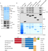
Glo3 forms a stoichiometric complex with COPI in vitro. (A) In vitro reconstitution of a COPI-Glo3 complex using TAP-purified coatomer, and recombinantly expressed and purified Glo3 lacking its distal amphipathic helix (459–493). Eluates obtained by Tobacco-etch virus (TEV) protease elution of TAP-tagged β′-COP after incubation with or without Glo3 (1–459) were analysed by Coomassie Blue staining of SDS-PAGE gels (top) or analysed by the immunodetection of western blots (bottom) using a coat antiserum detecting five of the seven subunits, or a Glo3 antibody. (B) Binding of coatomer to MBP fusion proteins of Glo3 from yeast lysates. The bound fraction was eluted and analysed by SDS-PAGE. Western blots were detected using a coat antiserum recognising five of the seven coatomer subunits. (C) Schematic illustration of the MBP-tagged truncations of Glo3. GAP, GTPase-activating domain; BoCCS, Binding of Coatomer, Cargo and SNARE domain; GRM, Glo3 regulatory motif; AmpH, amphipathic helix.
Fig. 3.
Fig. 3.
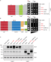
Gcs1 and Glo3 regulate distinct cellular functions. (A) Growth assay of Δgcs1 strains harbouring the indicated constructs on synthetic dropout medium analysed under MET25-promoter-repressing conditions (high methionine; control; indicated by a repressed promoter) or MET25-promoter-inducing conditions (normal methionine; test; indicated by an active promoter). A GAP-dead mutant of Gcs1 (Gcs1 R54K) supports growth at 30°C. Gcs1 L246D, ALPS mutant; Gcs1 AxxA, alanine for tryptophan substitution of the C-terminal tryptophan-based COPI recognition signal; Gcs1 Δ3xF AxxA, alanine substitution of the C-terminal tryptophan-based COPI recognition signal and three upstream phenylalanine residues. GAP, GTPase-activating domain; ALPS, ArfGAP1 lipid-packing sensor. (B) Growth assay of Δglo3 strains harbouring the indicated constructs on synthetic dropout medium was assayed under MET25 promoter-repressing conditions (high methionine; Control; indicated by a repressed promoter) and MET25-promoter-inducing conditions (normal methionine; test; indicated by an active promoter). A GAP-dead mutant of Glo3 (Glo3 R59K) does not support growth at 30°C, indicating that GTP hydrolysis stimulated by Glo3 in γ-Arf1 is essential. Glo3 ΔC, deletion of the amphipathic helix; Glo3 ΔGRM-ΔC, combined deletion of the amphipathic helix and the GRM; Glo3 Δ2x+ve, alanine substitution for the COPI-binding region. GAP, GTPase-activating domain; BoCCS, Binding of Coatomer, Cargo and SNARE domain; GRM, Glo3 regulatory motif; AmpH, amphipathic helix. (C) Expression analysis of proteins in the indicated strains grown under Met25-promoter-inducing conditions (normal methionine; test).
Fig. 4.
Fig. 4.
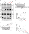
Snf1 phosphorylates Glo3 at S389. (A) Sequence alignment of the GRM domain of S. cerevisiae Glo3, and human ArfGAP2 and ArfGAP3. Asterisks indicate fully conserved residues; colons and full stops, respectively, indicate residues that are either strongly or weakly conserved. Regions of highest conservation – in comparison with other eukaryotes – are highlighted in orange. Residues S389 and S398 are indicated in green and purple, respectively. (B) Δglo3 strains expressing the indicated constructs under the control of a Tef1 promoter were analysed by Phos-tag PAGE. The electrophoretic mobility shift (arrowhead) indicates that residues S389 and S398 are phosphorylated in the presence and absence, respectively, of the Snf1 kinase. Treatment with λ-phosphatase (Ppase) resulted in dephosphorylation. (C) Analysis of proteins by Phos-tag PAGE and SDS-PAGE. Cells in mid-logarithmic phase were grown in the presence of glucose (+) or glucose starved (−) for 3 h prior to lysis. Western blots were immunodetected with the indicated antibodies. (D) Arf1-GFP fluorescence recovery after photobleaching (FRAP) in the presence (+D) or absence (−D) of glucose. Cells were grown to mid-logarithmic phase and glucose starved for 2 h prior to FRAP measurement. The plot, reflecting the recovery of Arf1-GFP at Golgi membranes (Vrg4-mCherry), shows the mean FRAP curves with the fits. The bar graphs on the right show Koff values per second (left) and Fm values in percent (right). (E) Arf1-GFP fluorescence recovery after photobleaching (FRAP) analysis in Δglo3 strains expressing the indicated Glo3 phospho-mutants. The plot, reflecting the recovery of Arf1-GFP at Golgi membranes (Vrg4-mCherry), shows the mean FRAP curves with the fits. The bar graphs on the right show Koff values per second (left) and Fm values in percent (right).
Fig. 5.
Fig. 5.
Phosphorylation and/or dephosphorylation regulates Glo3 function. (A) Growth assay of Δglo3 strains harbouring the indicated constructs on synthetic dropout medium analysed under MET25-promoter-repressing conditions (high methionine; control; indicated by a repressed promoter) and MET25-promoter-inducing conditions (normal methionine; test; indicated by an active promoter). The phosphomimetic mutant of the Glo3 motif rescues the dominant-negative effect of the GAP-dead Glo3 indicating that phosphorylation and/or dephosphorylation of the Glo3 motif regulates Glo3 function. (B) Expression analysis of proteins in the indicated strains grown under Met25-promoter-inducing conditions (normal methionine; test). (C) Affinity chromatography of GFP-tagged Glo3 variants from detergent extracts of Δglo3 strains harbouring the indicated GFP-tagged constructs and subsequent evaluation of COPI association. Glo3 ΔGRM-ΔC: combined deletion of amphipathic helix and Glo3 motif; Glo3 S389, 398A: alanine substitution for the indicated serines (non-phosphorylated mimetic); Glo3 S389, 398D: aspartic acid substitution for the indicated serine residues (phosphomimetic). Glo3 Δ2x+ve: alanine substitution for the COPI-binding region. (D) Summary. The Glo3 motif regulates the function of the GAP domain. Inactivation of the Glo3 motif by truncation or phosphorylation (phosphomimetic) rescues the dominant-negative lethality caused by a non-functional GAP domain. Inferred functionality of the GRM domain is highlighted in brown. GAP, GTPase activating domain; BoCCS, Binding of Coatomer, Cargo and SNARE domain; GRM, Glo3 regulatory motif; AmpH, amphipathic helix.
Fig. 6.
Fig. 6.
Manipulation of Gcs1 or COPI in the β-Arf niche has similar effects on cargo sorting. (A) Affinity chromatography of GFP-tagged Glo3 from detergent extracts of Δgcs1 strains harbouring the indicated Gcs1 constructs and subsequent evaluation of COPI and Gcs1 association. Gcs1 R54K, GAP-dead mutant of Gcs1; L246D, ALPS mutant; Gcs1 AxxA, alanine for tryptophan substitution of the C-terminal tryptophan-based COPI recognition signal. (B) Secretion assay of the indicated strains harbouring the indicated plasmids. Proteins secreted into the culture medium were analysed by SDS-PAGE and immunoblot analysis using antibodies specific for Pdi1. Cell pellets from the same cultures were also analysed using antibodies specific for Pgk1 and Gcs1. (C) Plot of the relative amounts of Pdi1 secreted into the culture medium by the indicated strains, as quantified by the densiometric analysis of immunoblots. Quantification of three independent experiments. Error bars depict ±s.e.m. (D) Secretion assay of the indicated strains harbouring the indicated plasmids. Proteins secreted into the culture medium were analysed by SDS-PAGE and immunoblot analysis using antibodies specific for Pdi1. Cell pellets from the same cultures were also analysed using antibodies specific for Pgk1 and Glo3. (E) Plot of the relative amounts of Pdi1 secreted into the culture medium by the indicated strains, as quantified by the densiometric analysis of immunoblots. Quantification of four independent experiments. Error bars indicate±s.e.m.
Fig. 7.
Fig. 7.
The GRM domain regulates Glo3 function. (A) Summary of the key results obtained by manipulating the GAP and the GRM domain. (B) Schematic illustration of the COPI triad for structural orientation. (C) Adjacent GRM domains interconnect individual coat molecules at the heart of the COPI triad, effectively locking the triad together and stabilising COPI on the membrane. GTP hydrolysis in γ-Arf triggers a conformational change in the GRM domain and (D) uncouples adjacent GRM domains, effectively unlocking the triad and promoting the dissociation of COPI. (E) In this depiction, the GRM domain interconnects adjacent coat heptamers within a triad rather than adjacent GRM domains of neighbouring Glo3 molecules.

References

    1. Aguilera-Romero A., Kaminska J., Spang A., Riezman H. and Muñiz M. (2008). The yeast p24 complex is required for the formation of COPI retrograde transport vesicles from the Golgi apparatus. J. Cell Biol. 180, 713-720. 10.1083/jcb.200710025 - DOI - PMC - PubMed
    1. Alisaraie L. and Rouiller I. (2012). Full-length structural model of RET3 and SEC21 in COPI: identification of binding sites on the appendage for accessory protein recruitment motifs. J. Mol. Model. 18, 3199-3212. 10.1007/s00894-011-1324-9 - DOI - PMC - PubMed
    1. Aniento F., Gu F., Parton R. G. and Gruenberg J. (1996). An endosomal beta COP is involved in the pH-dependent formation of transport vesicles destined for late endosomes. J. Cell Biol. 133, 29-41. 10.1083/jcb.133.1.29 - DOI - PMC - PubMed
    1. Antonny B., Beraud-Dufour S., Chardin P. and Chabre M. (1997). N-terminal hydrophobic residues of the G-protein ADP-ribosylation factor-1 insert into membrane phospholipids upon GDP to GTP exchange. Biochemistry 36, 4675-4684. 10.1021/bi962252b - DOI - PubMed
    1. Antonny B., Madden D., Hamamoto S., Orci L. and Schekman R. (2001). Dynamics of the COPII coat with GTP and stable analogues. Nat. Cell Biol. 3, 531-537. 10.1038/35078500 - DOI - PubMed

Publication types

MeSH terms

Substances