Review
. 2019 Jul 22;146(14):dev167700.
doi: 10.1242/dev.167700.

Model systems for regeneration: salamanders

Affiliations
Review

Model systems for regeneration: salamanders

Alberto Joven et al. Development. .

Abstract

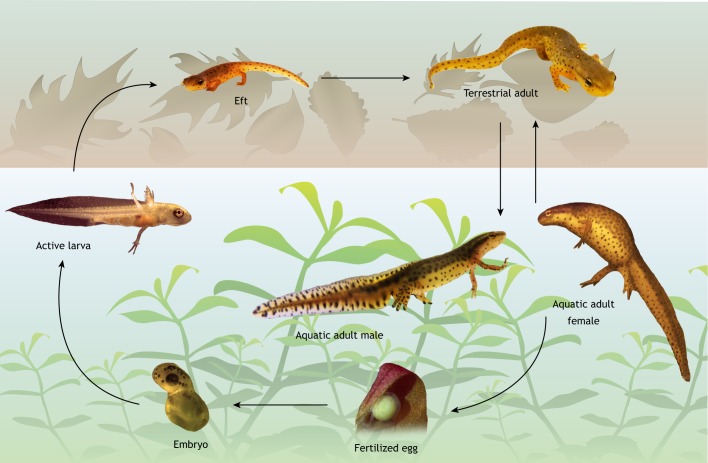
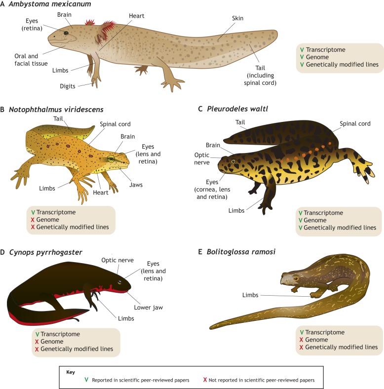
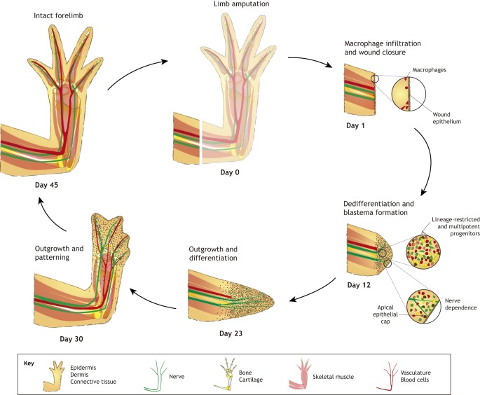
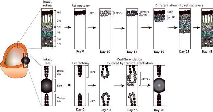
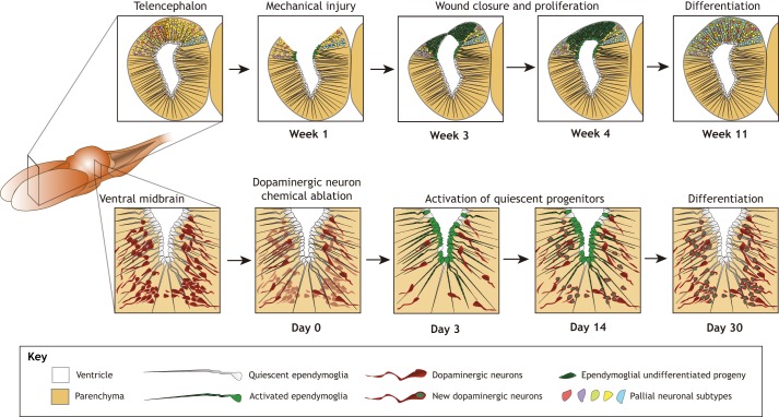
Salamanders have been hailed as champions of regeneration, exhibiting a remarkable ability to regrow tissues, organs and even whole body parts, e.g. their limbs. As such, salamanders have provided key insights into the mechanisms by which cells, tissues and organs sense and regenerate missing or damaged parts. In this Primer, we cover the evolutionary context in which salamanders emerged. We outline the varieties of mechanisms deployed during salamander regeneration, and discuss how these mechanisms are currently being explored and how they have advanced our understanding of animal regeneration. We also present arguments about why it is important to study closely related species in regeneration research.

Keywords: Axolotl; CNS; Genome; Limb; Model organism; Newt.


Conflict of interest statement

Competing interestsThe authors declare no competing or financial interests.

Figures

Fig. 1.
Fig. 1.
Salamanders display complex life cycles in both terrestrial and aquatic habitats. A typical salamander life cycle (exemplified here by that of Notophthalmus viridescens) involves both terrestrial and aquatic stages. Adult newts alternate facultative aquatic/terrestrial lifestyles, but they mate and lay fertilized eggs in the water. These eggs then develop into embryos that hatch as aquatic larvae. The larvae are ferocious zooplankton hunters that undergo metamorphosis prior to leaving the aquatic milieu and becoming terrestrial juveniles (termed efts), which seasonally return to water to breed after they reach sexual maturity. Many salamander species are entirely land living without an aquatic larval stage.
Fig. 2.
Fig. 2.
Salamander species in regeneration research. Both established and emerging species are shown, highlighting regenerative organs/tissues and major resources available for each species. (A) The Mexican axolotl, Ambystoma mexicanum, is a paedomorphic salamander that retains fully aquatic features throughout its entire life cycle. The axolotl is easy to breed under laboratory conditions, and is the most commonly used salamander model organism in regeneration research, mostly owing to the availability of several genetically modified lines (Tanaka, 2016). (B) The Eastern red spotted newt, Notophthalmus viridescens, has contributed significantly to our understanding of multiple regeneration processes with reference transcriptomes available. Genetically modified lines are difficult to establish due to its long generation time and complex life cycle (Abdullayev et al., 2013; Looso et al., 2013). (C) The Iberian ribbed newt, Pleurodeles waltl, is a highly regenerative, emerging model species. It can be maintained in a fully aquatic habitat throughout its entire life cycle and has a similar generation time to the axolotl. Transcriptomes and genome assemblies are now available, as well as genetically modified lines (Elewa et al., 2017; Hayashi and Takeuchi, 2015; Hayashi et al., 2013; Joven et al., 2015, 2018). (D) The Japanese fire-bellied newt, Cynops pyrrhogaster, has been used to study eye, limb, jaw and brain regeneration. A transcriptome focusing on lens and neural retina regeneration has been assembled (Casco-Robles et al., 2016; Kurosaka et al., 2008; Nakamura et al., 2014). (E) The plethodontid Bolitoglossa ramosi is a fully terrestrial, direct developer (no larval stage) for which a limb regeneration transcriptome has been reported (Arenas Gomez et al., 2017, 2018).
Fig. 3.
Fig. 3.
Key processes during limb regeneration. The salamander limb contains all typical structural elements of tetrapods. Upon amputation, salamander limb regeneration starts by scar-free wound healing and wound closure. Infiltrating macrophages are essential for this event, probably for clearing debris, although other signalling mechanisms cannot be excluded. Cells in the mature limb then undergo reprogramming/dedifferentiation to form a blastema. The degree of reprogramming varies between cell types and species. Nerve-derived factors are required for subsequent blastema cell proliferation and outgrowth. The cells also retain positional memory during the regeneration event, allowing them to undergo the appropriate patterning to re-from an intact limb. The time course of regeneration indicated in this figure is based on staging in adult Notophthalmus viridescens.
Fig. 4.
Fig. 4.
Regeneration of ocular tissues. (Top) Following retinectomy (detachment of the RPE from the photoreceptor cell layer), a new pigmented cell layer appears first. This is followed by the formation of neuro-retinal cell types in an order that recapitulates development: ganglion cells form first, followed by amacrine cells, horizontal cells and Müller glia. (Bottom) The lens can regenerate following lentectomy (lens removal), via the dedifferentiation and subsequent transdifferentiation of pigmented epithelial cells of the dorsal iris. Newts retain lens regeneration ability throughout adulthood, unlike axolotls, in which the ability to regenerate the lens is lost 2 weeks after hatching. dIPE, dorsal iris pigmented epithelium; dIPESCs, dorsal iris pigmented epithelium cells; GCL, ganglion cell layer; INL, inner nuclear layer; ONL, outer nuclear layer; OPL, outer plexiform layer; proNR, inner rudimentary layer; proRE, retinal pigmented epithelium rudimentary layer; RPE, retinal pigmented epithelium; RPESCs, retinal pigmented epithelium stem cells; vIPE, ventral iris pigmented epithelium.
Fig. 5.
Fig. 5.
Brain regeneration in salamanders. (Top) Brain regeneration following injury (e.g. unilateral forebrain extirpation) presumably occurs by activation of ependymoglial cells. Neuronal diversity is restored but projections are not a faithful replication of the original. The time frame shown here is based on studies in the axolotl. (Bottom) Studies on red-spotted newts have showed that dopaminergic and cholinergic (not shown) neurons regenerate in several brain regions after the selective ablation of individual neuronal subtypes. Injury-responsive neurogenesis is fuelled by reactivation of quiescent resident neuronal progenitor cells, the ependymoglial cells, which are the equivalent of glial cells in mammals.

References

    1. Abdullayev I., Kirkham M., Björklund A. K., Simon A. and Sandberg R. (2013). A reference transcriptome and inferred proteome for the salamander Notophthalmus viridescens. Exp. Cell Res. 319, 1187-1197. 10.1016/j.yexcr.2013.02.013 - DOI - PubMed
    1. Amamoto R., Huerta V. G., Takahashi E., Dai G., Grant A. K., Fu Z. and Arlotta P. (2016). Adult axolotls can regenerate original neuronal diversity in response to brain injury. Elife 5, e13998 10.7554/eLife.13998 - DOI - PMC - PubMed
    1. Anokye-Danso F., Trivedi C. M., Juhr D., Gupta M., Cui Z., Tian Y., Zhang Y., Yang W., Gruber P. J., Epstein J. A. et al. (2011). Highly efficient miRNA-mediated reprogramming of mouse and human somatic cells to pluripotency. Cell Stem Cell 8, 376-388. 10.1016/j.stem.2011.03.001 - DOI - PMC - PubMed
    1. Arenas Gomez C. M., Gomez Molina A., Zapata J. D. and Delgado J. P. (2017). Limb regeneration in a direct-developing terrestrial salamander, Bolitoglossa ramosi (Caudata: Plethodontidae): limb regeneration in plethodontid salamanders. Regeneration (Oxf) 4, 227-235. 10.1002/reg2.93 - DOI - PMC - PubMed
    1. Arenas Gomez C. M., Woodcock R. M., Smith J. J., Voss R. S. and Delgado J. P. (2018). Using transcriptomics to enable a plethodontid salamander (Bolitoglossa ramosi) for limb regeneration research. BMC Genomics 19, 704 10.1186/s12864-018-5076-0 - DOI - PMC - PubMed

Publication types