. 2019 Oct;18(10):1950-1966.
doi: 10.1074/mcp.RA119.001356. Epub 2019 Jul 22.

A Systems-level Characterization of the Differentiation of Human Embryonic Stem Cells into Mesenchymal Stem Cells

Affiliations

A Systems-level Characterization of the Differentiation of Human Embryonic Stem Cells into Mesenchymal Stem Cells

Anja M Billing et al. Mol Cell Proteomics. 2019 Oct.

Abstract

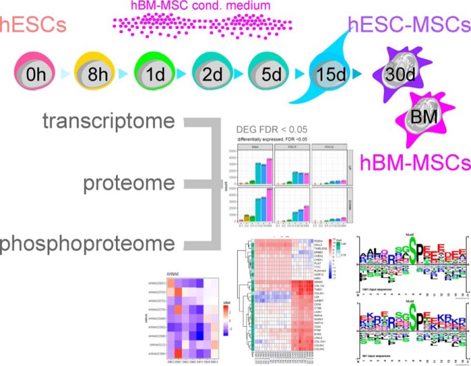
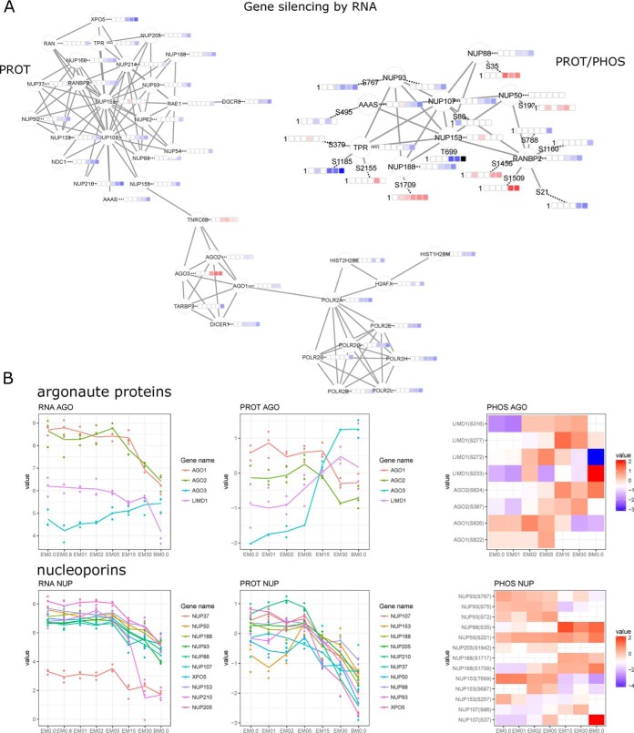
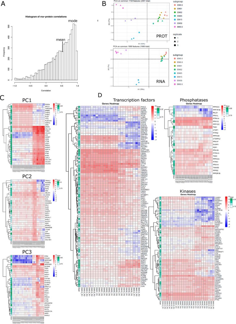
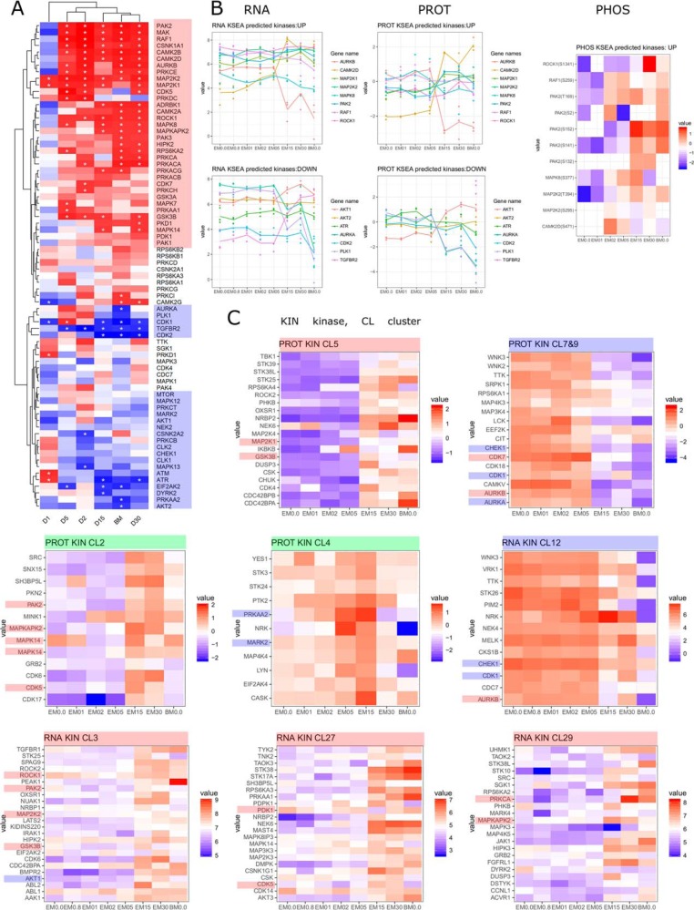
Mesenchymal stem/stromal cells (MSCs) are self-renewing multipotent cells with regenerative, secretory and immunomodulatory capabilities that are beneficial for the treatment of various diseases. To avoid the issues that come with using tissue-derived MSCs in therapy, MSCs may be generated by the differentiation of human embryonic stems cells (hESCs) in culture. However, the changes that occur during the differentiation process have not been comprehensively characterized. Here, we combined transcriptome, proteome and phosphoproteome profiling to perform an in-depth, multi-omics study of the hESCs-to-MSCs differentiation process. Based on RNA-to-protein correlation, we determined a set of high confidence genes that are important to differentiation. Among the earliest and strongest induced proteins with extensive differential phosphorylation was AHNAK, which we hypothesized to be a defining factor in MSC biology. We observed two distinct expression waves of developmental HOX genes and an AGO2-to-AGO3 switch in gene silencing. Exploring the kinetic of noncoding ORFs during differentiation, we mapped new functions to well annotated long noncoding RNAs (CARMN, MALAT, NEAT1, LINC00152) as well as new candidates which we identified to be important to the differentiation process. Phosphoproteome analysis revealed ESC and MSC-specific phosphorylation motifs with PAK2 and RAF1 as top predicted upstream kinases in MSCs. Our data represent a rich systems-level resource on ESC-to-MSC differentiation that will be useful for the study of stem cell biology.

Keywords: LC-MS/MS; RNA SEQ; cell differentiation; developmental biology; differentiation; gene expression; human embryonic stem cells; human mesenchymal stem cells; phosphoproteome; post-translational modifications; quantification; quantitative proteomics; stem cells; systems biology.


Conflict of interest statement

The authors declare no competing interests

Figures

None
Graphical abstract
Fig. 1.
Fig. 1.
ESC to MSC differentiation. A, Schematic of the differentiation protocol with sampling schedule for the different techniques. B, Summary of omics technologies and their analytical depth to profile the differentiation from ESCs to MSCs. C, Expression profiles of selected markers for ESCs, MSCs, and (D) mesoderm, as well as (E) RNA transcripts upregulated during differentiation from 8 h onwards.
Fig. 2.
Fig. 2.
Differential expression analysis. A, Differentially expressed features for each technique. B, Enrichment analysis on differentially expressed features (FDR < 0.05) of RNA, PROT and PHOS. Tile plots are shown for selected enriched GOBP terms. C, Expression profiles of selected lncRNA and antisense RNA transcripts.
Fig. 3.
Fig. 3.
Gene silencing by RNA during ESC to MSC differentiation. A, Protein-protein interaction network for proteome and phosphoproteome data. B, Expression (RNA/PROT) and phosphorylation profiles (PHOS) for argonaute proteins and nucleoporins.
Fig. 4.
Fig. 4.
Fuzzy c-means clustering on differentially expressed features for each technique. Data was partitioned in either 32 (RNA) or 16 clusters (PROT, PHOS). A, PCA of clusters for each technique. Clusters with exceptional high increase (red), decrease (blue) or peak expression at intermediate differentiation stage (D15) (green) are highlighted. B, Expression profiles of selected RNA and PROT clusters. C, Selected clusters were filtered for transcription factors. D, Quantified and differentially expressed (DEGs) features per technique were filtered for transcription factors (TF), kinases (KIN) and phosphatases (PPT).
Fig. 5.
Fig. 5.
RNA-PROT correlation. A, Distribution of RNA-PROT correlation with mean and mode indicated. B, PCA on features with R > 0.7 and FDR < 0.05 for PROT and RNA. C, Hierarchical clustering of top30 features per principal components 1–3. D, Hierarchical clustering of transcription factors, kinases and phosphatases, which show good RNA-PROT correlation (R > 0.7, FDR < 0.05). RNA transcripts are shown with RNA-PROT correlation indicated on the left. Legends for expression intensity and correlation are on the right.
Fig. 6.
Fig. 6.
Phosphoproteome profiling - phosphomotif analysis. A, Class I phosphosites in our data set (PHOS) compared with the whole human PhosphoSitePlus database (PPSP) or entries for hESCs (PPSP_ESC) and hMSCs (PPSP_MSC). B, Top10 phosphoproteins with the most phosphosites per protein. Half of them displayed intense phosphorylation during differentiation (*). C, Expression profiles for top phosphoproteins. D, Differential phosphorylation for AHNAK. E, Counts of enriched phosphomotifs with reduced (left) or induced (right) phosphorylation during ESC to MSC differentiation. ESC or MSC specific phosphomotifs common for D15, D30, BM. F, Combined sequence logo for phosphomotifs with reduced phosphorylation (ESCs) or increased phosphorylation (MSCs) during differentiation.
Fig. 7.
Fig. 7.
Kinases during differentiation. A, Upstream kinase prediction by KSEA. B, Expression profiles of KSEA predicted kinases at RNA, protein and phosphoprotein level. C, Expression profiles of selected clusters filtered for kinases which show distinct induction (red), reduction (blue) or peak expression (green). Predicted kinases are highlighted according to their predicted activity.

References

    1. Wei X., Yang X., Han Z., Qu F., Shao L., and Shi Y. (2013) Mesenchymal stem cells: a new trend for cell therapy. Acta Pharmacol. Sin. 34, 747–754 - PMC - PubMed
    1. Wagner W., Bork S., Horn P., Krunic D., Walenda T., Diehlmann A., Benes V., Blake J., Huber F.-X., Eckstein V., Boukamp P., and Ho A. D. (2009) Aging and replicative senescence have related effects on human stem and progenitor cells. PLoS ONE 4, e5846. - PMC - PubMed
    1. Hematti P. (2011) Human embryonic stem cell–derived mesenchymal stromal cells. Transfusion 51, 138S–144S - PMC - PubMed
    1. Arpornmaeklong P., Brown S. E., Wang Z., and Krebsbach P. H. (2009) Phenotypic characterization, osteoblastic differentiation, and bone regeneration capacity of human embryonic stem cell–derived mesenchymal stem cells. Stem Cells Dev. 18, 955–968 - PMC - PubMed
    1. Barberi T., Willis L. M., Socci N. D., and Studer L. (2005) Derivation of multipotent mesenchymal precursors from human embryonic stem cells. PLoS Med. 2, e161. - PMC - PubMed

Publication types