. 2019 Jul 22;9(1):10564.
doi: 10.1038/s41598-019-46661-3.

Surgical anatomy of the ovine sural nerve for facial nerve regeneration and reconstruction research

Affiliations

Surgical anatomy of the ovine sural nerve for facial nerve regeneration and reconstruction research

Yosuke Niimi et al. Sci Rep. .

Abstract

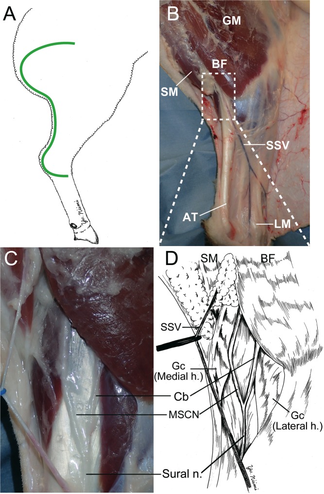
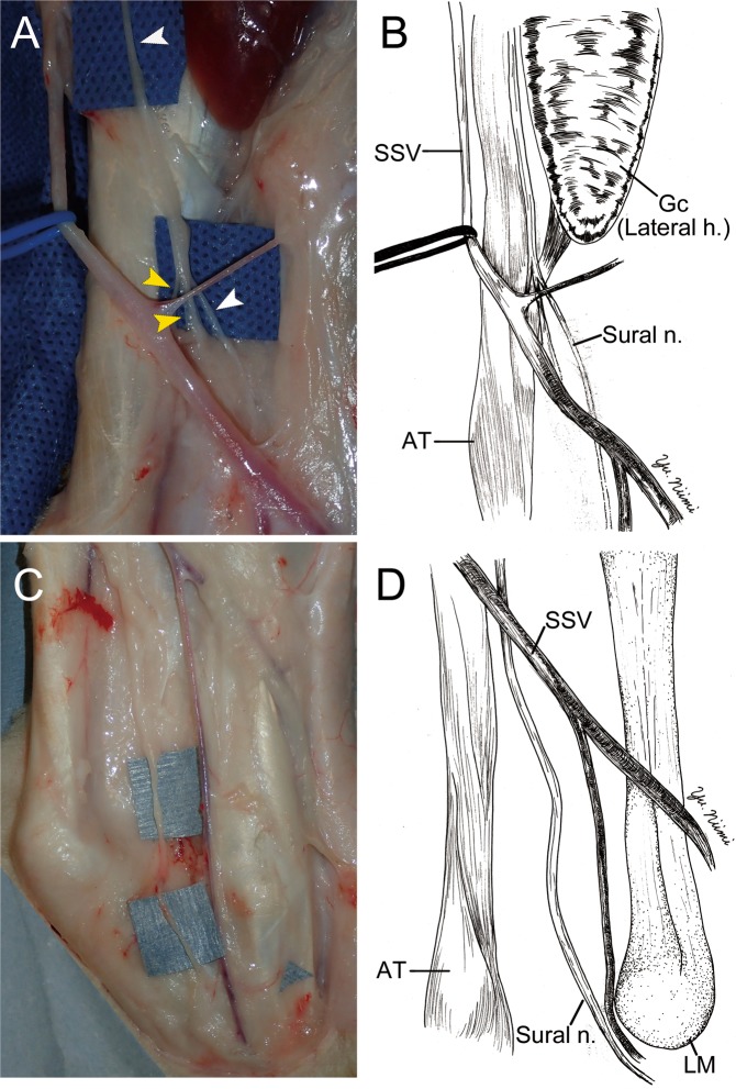
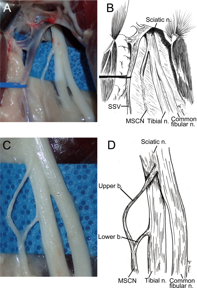
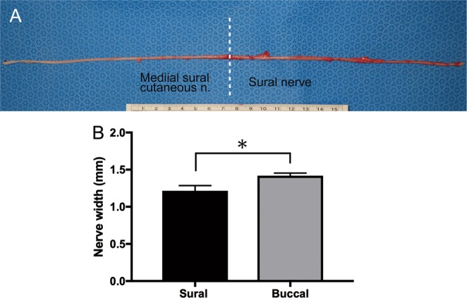
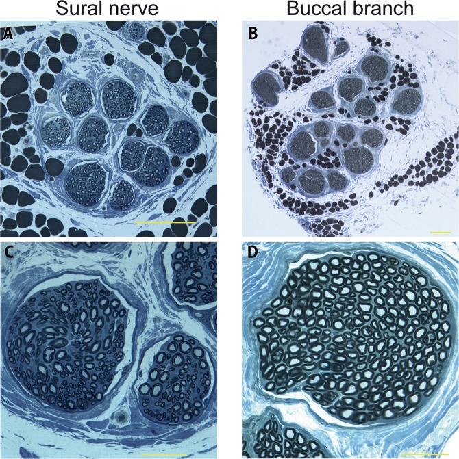
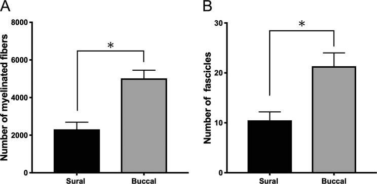
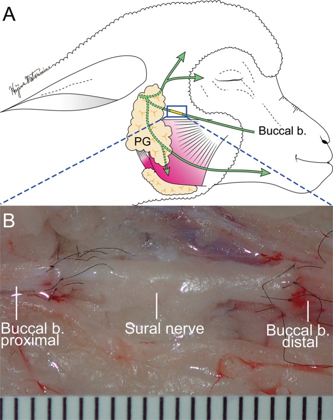
The lack of a clinically relevant animal models for research in facial nerve reconstruction is challenging. In this study, we investigated the surgical anatomy of the ovine sural nerve as a potential candidate for facial nerve reconstruction, and performed its histological quantitative analysis in comparison to the buccal branch (BB) of the facial nerve using cadaver and anesthetized sheep. The ovine sural nerve descended to the lower leg along the short saphenous vein. The length of the sural nerve was 14.3 ± 0.5 cm. The distance from the posterior edge of the lateral malleolus to the sural nerve was 7.8 ± 1.8 mm. The mean number of myelinated fibers in the sural nerve was significantly lower than that of the BB (2,311 ± 381vs. 5,022 ± 433, respectively. p = 0.003). The number of fascicles in the sural nerve was also significantly lower than in the BB (10.5 ± 1.7 vs. 21.3 ± 2.7, respectively. p = 0.007). The sural nerve was grafted to the BB with end-to-end neurorrhaphy under surgical microscopy in cadaver sheep. The surgical anatomy and the number of fascicles of the ovine sural nerve were similar of those reported in humans. The results suggest that the sural nerve can be successfully used for facial nerve reconstruction research in a clinically relevant ovine model.


Conflict of interest statement

The authors declare no competing interests.

Figures

Figure 1
Figure 1
Surgical procedure to identify of the ovine sural nerve. (A) The green line shows the incision line. From the lateral gluteal-thigh border to 2 cm posterior the lateral malleolus C-shape incision was made on the ovine cadaver right leg. (B) The gluteus maximus (GM), biceps femoris (BF), semimembranosus (SM), Achilles tendon (AT), lateral malleolus (LM) were dissected after undermining the skin flap. The short saphenous vein (SSV) was identified. (C,D) The sural nerve, medial sural cutaneous nerve (MSCN), and communicating branch (Cb) were identified to be located under the gastrocnemius fascia, between the lateral and medial head of the gastrocnemius, and distal of BF. Sural nerve formed by MSCN and Cb. Gc (Medial h.): Gastrocnemius medial head. Gc (Lateral h.): Gastrocnemius lateral head.
Figure 2
Figure 2
The surgical anatomy of sural nerve (Sural n.); middle (A,B) and distal (C,D) portion of the leg. (A,B) The sural nerve (white arrow head) ran parallel to the short saphenous vein (SSV, blue rubber tape), and ran medial side of the gastrocnemius (Gc) lateral head (Lateral h.). Thereafter, two nerves (yellow arrow heads) were merging to the sural nerve at the midpoint of the lower leg. (C,D) In the distal portion of lower leg, the sural nerve was passed posterior to the lateral malleolus (LM). Thereafter, the sural nerve penetrated the deep fascia and went into the foot area. AT: Achilles tendon. Gc (Lateral h.): Gastrocnemius lateral head.
Figure 3
Figure 3
The surgical anatomy of medial sural cutaneous nerve (MSCN). (A,B) To dissect to the proximal region from the point of Fig. 1, MSCN was divided from sciatic nerve (Sciatic n.) near the tibial nerve side between biceps femoris and semimembranosus. Common fibular nerve (Common fibular n.) was also devided from scaiatic nerve at the same place. (C,D) After removing connective tissue, MSCN was exposed forming from two branches, which were originated from sciatic and tibial nerves.
Figure 4
Figure 4
(A) The picture of sural nerve and medial sural cutaneous nerve. The average lengths of the medial sural cutaneous and sural nerve were 14.3 ± 0.5 (n = 7) and 16.4 ± 0.8 cm (n = 7), respectively. The average of total length was 30.2 ± 0.7 cm (n = 16). Unit of the ruler: cm (B) The width of sural nerve (n = 10) and buccal branch (n = 8) were measured using the photographs by ImageJ software version 1.50 (National Institutes of Health, Bethesda, MD) The width of the sural nerve was significantly lower than that of the buccal branch (1.2 ± 0.07 vs. 1.4 ± 0.04 mm respectively. p = 0.0117).
Figure 5
Figure 5
The toluidine blue stain of sural nerve (Left column) and buccal branch (right column). Both sural nerve (A) and buccal branch (B) had epineurium. (C) Myelin sheaths of axons of the sural nerve was stained blue color and observed clearly. (D) Dense myelin sheath of buccal branch was observed. (A,B) Scale bar: 200 μm. (C,D) Scale bar: 50 μm.
Figure 6
Figure 6
Histological assessment. (A) The mean number of myelinated fibers in the Sural nerve were significantly lower than that in the buccal branch (2,311 ± 381 vs. 5,022 ± 433, respectively, p = 0.003). (B) The number of fascicles in the sural nerve were significantly lower than that in the buccal branch (10.5 ± 1.7 vs. 21.3 ± 2.7, respectively, p = 0.007).
Figure 7
Figure 7
Demonstration of sural nerve graft to the buccal branch with end-to-end neurorrhaphy. (A) Schematic diagram of sural nerve graft. end-to-end neurorrhaphy was performed 10 mm distal to the edge of parotid gland (PG). (B) Microscopical image of end-to-end neurorrhaphy. Fifteen millimeter-sural nerve was grafted 6 perineural sutures using 10-0 nylon to bridge the defect of buccal branch with end-to-end neurorrhaphy under the surgical microscope. One unit indicates 1 mm.

Similar articles

Cited by

References

    1. Jackson CG, Glasscock ME, III, Hughes G, Sismanis A. Facial paralysis of neoplastic origin: diagnosis and management. Laryngoscope. 1980;90:1581–1595. doi: 10.1288/00005537-198010000-00001. - DOI - PubMed
    1. Terzis JK, Sun DD, Thanos PK. Historical and basic science review: Past present, and future of nerve repair. Journal of reconstructive microsurgery. 1997;13:215–225. doi: 10.1055/s-2007-1006407. - DOI - PubMed
    1. Lee EI, Hurvitz KA, Evans GRD, Wirth GA. Cross-facial nerve graft: past and present. Journal of Plastic, Reconstructive & Aesthetic Surgery. 2007;61:250–256. doi: 10.1016/j.bjps.2007.05.016. - DOI - PubMed
    1. Matsumine H, Sasaki R, Yamato M, Okano T, Sakurai H. A polylactic acid non-woven nerve conduit for facial nerve regeneration in rats. J Tissue Eng Regen Med. 2014;8:454–462. doi: 10.1002/term.1540. - DOI - PubMed
    1. Niimi Y, et al. A collagen-coated PGA conduit for interpositional-jump graft with end-to-side neurorrhaphy for treating facial nerve paralysis in rat. Microsurgery. 2019;39:70–80. doi: 10.1002/micr.30291. - DOI - PubMed

Publication types