

Three-dimensional HepaRG spheroids as a liver model to study human genotoxicity in vitro with the single cell gel electrophoresis assay
- PMID: 31332230
- PMCID: PMC6646340
- DOI: 10.1038/s41598-019-47114-7
Three-dimensional HepaRG spheroids as a liver model to study human genotoxicity in vitro with the single cell gel electrophoresis assay
Abstract
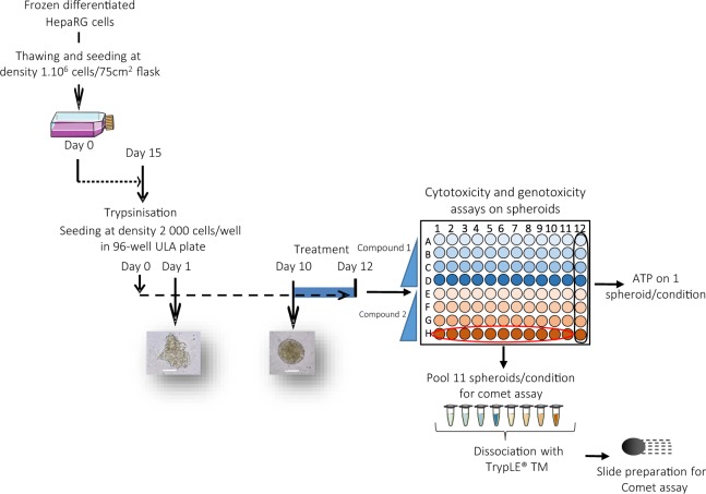
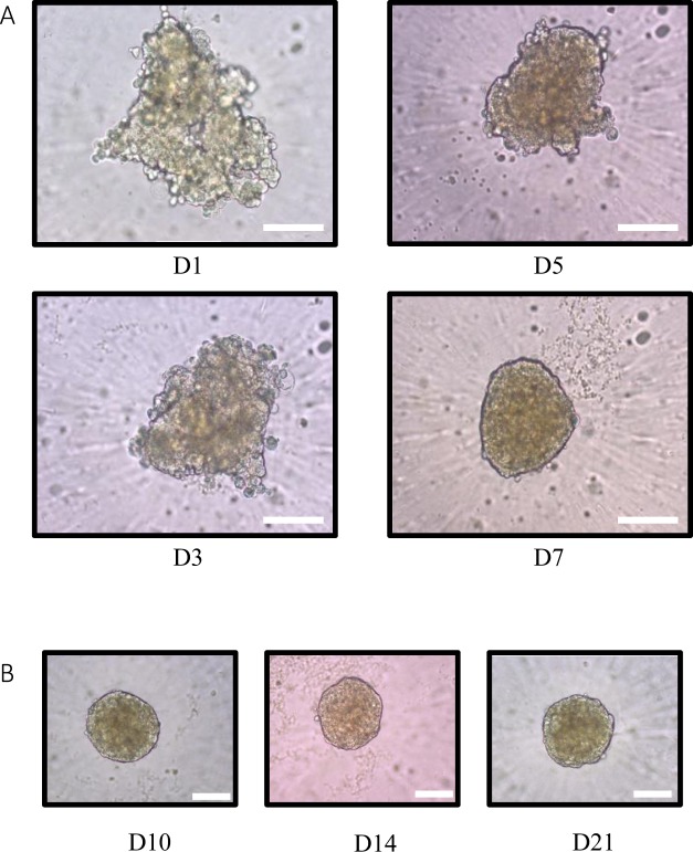
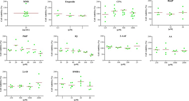
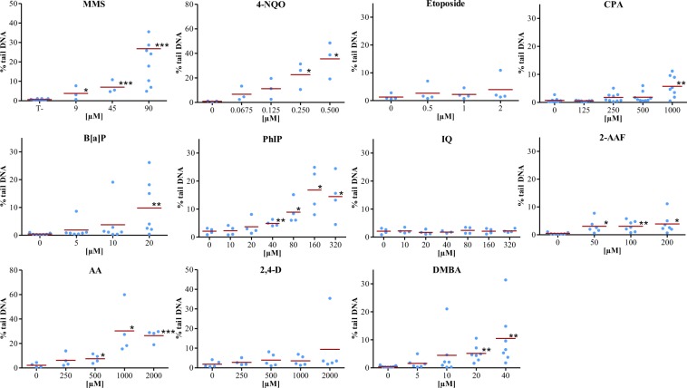
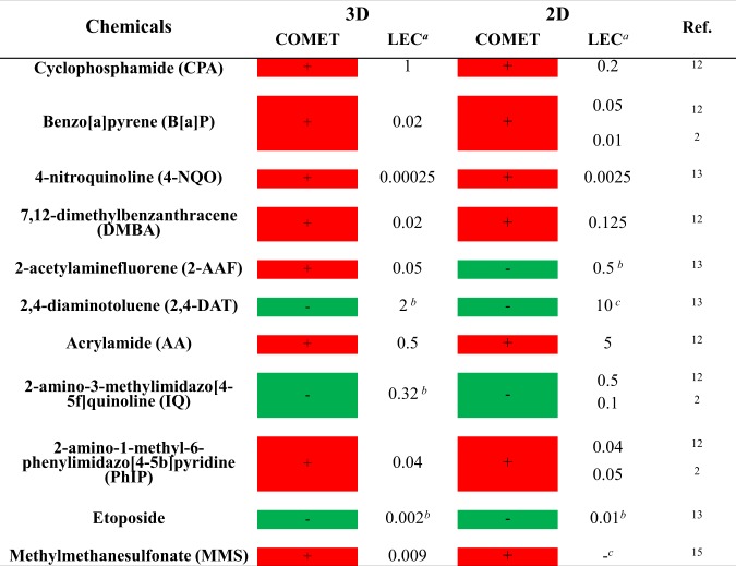
Many efforts have been made in the last 30 years to develop more relevant in vitro models to study genotoxic responses of drugs and environmental contaminants. While 2D HepaRG cells are one of the most promising models for liver toxicology, a switch to 3D cultures that integrate both in vivo architecture and cell-cell interactions has occurred to achieve even more predictive models. Preliminary studies have indicated that 3D HepaRG cells are suitable for liver toxicity screening. Our study aimed to evaluate the response of HepaRG spheroids exposed to various genotoxic compounds using the single cell gel electrophoresis assay. HepaRG spheroids were used at 10 days after seeding and exposed for 24 and 48 hours to certain selected chemical compounds (methylmethansulfonate (MMS), etoposide, benzo[a]pyrene (B[a]P), cyclophosphamide (CPA), 7,12-dimethylbenz[a]anthracene (DMBA), 2-acetylaminofluorene (2-AAF), 4-nitroquinoline (4-NQO), 2-amino-1-methyl-6-phenylimidazo[4,5-b]pyridine (PhIP), 2-amino-3-methylimidazo[4,5-f]quinolone (IQ), acrylamide, and 2-4-diaminotoluene (2,4-DAT)). After treatment, the comet assay was performed on single cell suspensions and cytotoxicity was determined by the ATP assay. Comet formation was observed for all compounds except IQ, etoposide and 2,4-DAT. Treatment of spheroids with rifampicin increased CYP3A4 activity, demonstrating the metabolic capacity of HepaRG spheroids. These data on genotoxicity in 3D HepaRG spheroids are promising, but further experiments are required to prove that this model can improve the predictivity of in vitro models to detect human carcinogens.
Conflict of interest statement
The authors declare no competing interests.
Figures
References
-
- Godoy P, et al. Recent advances in 2D and 3D in vitro systems using primary hepatocytes, alternative hepatocyte sources and non-parenchymal liver cells and their use in investigating mechanisms of hepatotoxicity, cell signaling and ADME. Archives of Toxicology. 2013;87:1315–1530. doi: 10.1007/s00204-013-1078-5. - DOI - PMC - PubMed
MeSH terms
Substances
LinkOut - more resources
Full Text Sources
Other Literature Sources
Miscellaneous
