. 2019 Jul 22;9(1):10568.
doi: 10.1038/s41598-019-47122-7.

Monte-Carlo dosimetry and real-time imaging of targeted irradiation consequences in 2-cell stage Caenorhabditis elegans embryo

Affiliations

Monte-Carlo dosimetry and real-time imaging of targeted irradiation consequences in 2-cell stage Caenorhabditis elegans embryo

Eva Torfeh et al. Sci Rep. .

Abstract

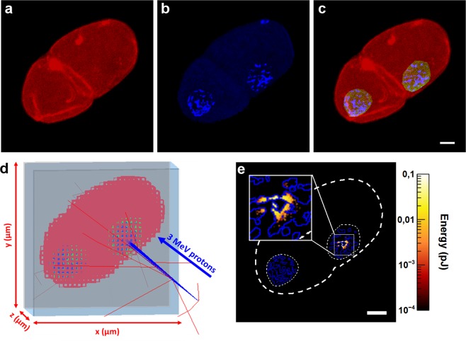
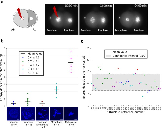
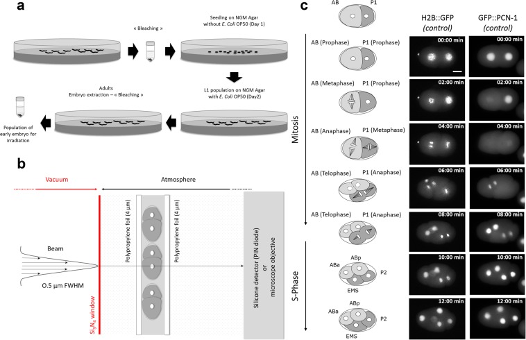
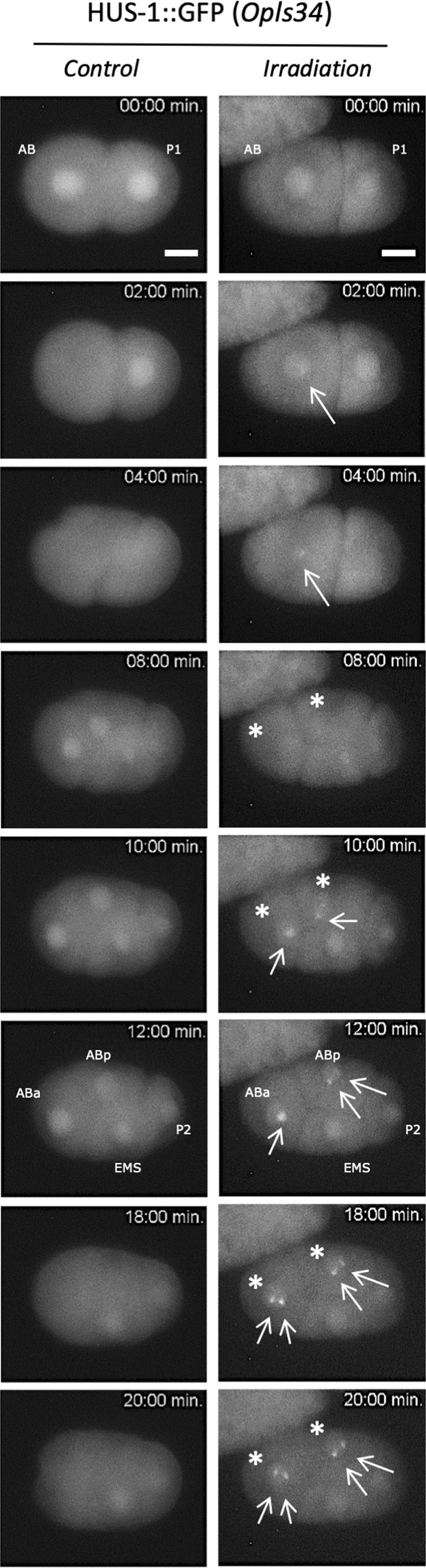
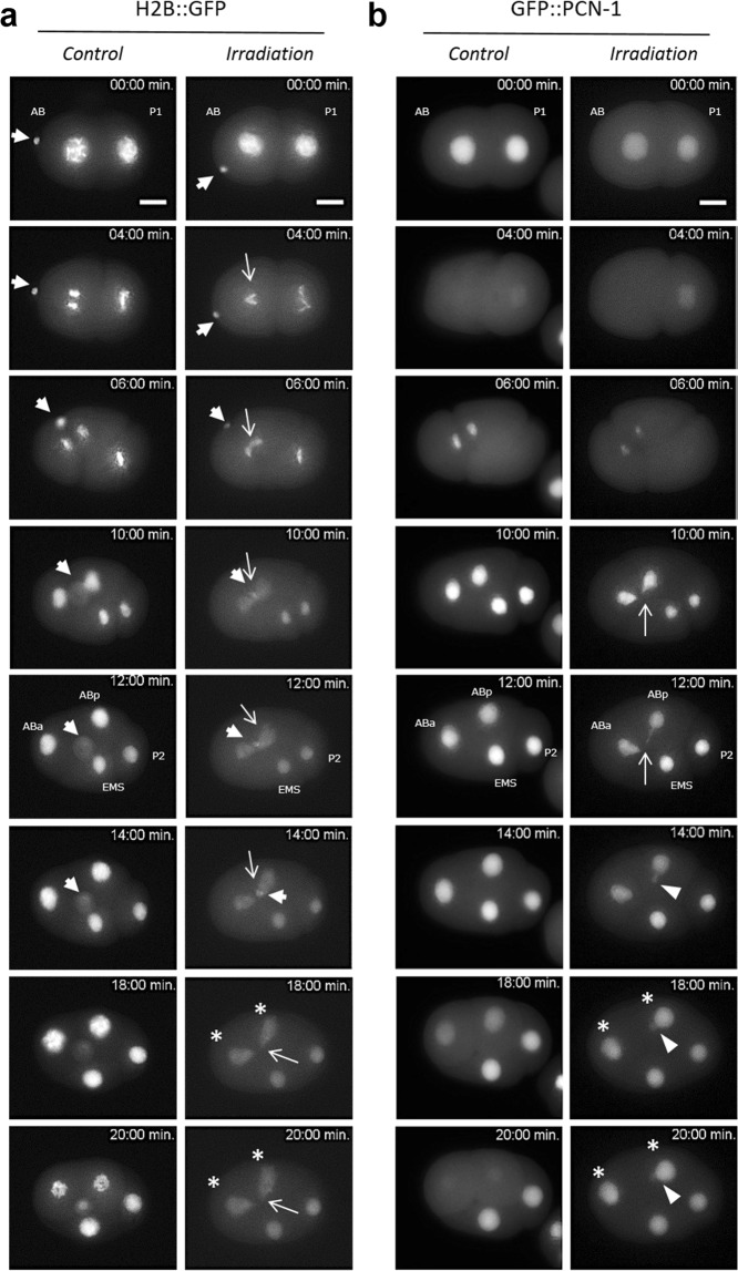
Charged-particle microbeams (CPMs) provide a unique opportunity to investigate the effects of ionizing radiation on living biological specimens with a precise control of the delivered dose, i.e. the number of particles per cell. We describe a methodology to manipulate and micro-irradiate early stage C. elegans embryos at a specific phase of the cell division and with a controlled dose using a CPM. To validate this approach, we observe the radiation-induced damage, such as reduced cell mobility, incomplete cell division and the appearance of chromatin bridges during embryo development, in different strains expressing GFP-tagged proteins in situ after irradiation. In addition, as the dosimetry of such experiments cannot be extrapolated from random irradiations of cell populations, realistic three-dimensional models of 2 cell-stage embryo were imported into the Geant4 Monte-Carlo simulation toolkit. Using this method, we investigate the energy deposit in various chromatin condensation states during the cell division phases. The experimental approach coupled to Monte-Carlo simulations provides a way to selectively irradiate a single cell in a rapidly dividing multicellular model with a reproducible dose. This method opens the way to dose-effect investigations following targeted irradiation.


Conflict of interest statement

The authors declare no competing interests.

Figures

Figure 1
Figure 1
Monte-Carlo dosimetry and calculation of the energy deposit in 2-cell stage C. elegans embryo. (a,b) Confocal images of a 2-cell stage embryo stained with phalloidin (red) and Hoechst33342 (blue). (c) The chromatin (blue) and the embryo (red) are identified by applying an intensity threshold to separate fluorescent objects (chromatin) from the background. The nuclear volume (green) is outlined manually. (d) Voxelized geometry in Geant4 (low resolution) irradiated with 3 MeV protons. The incident protons are represented by blue lines, the red lines illustrate the trajectory of secondary electrons generated in air. (e) Z-projection of the energy deposit calculated using Geant4-DNA after irradiation with 103 protons. The dashed outline corresponds to the contouring projection of the 3 volumes (chromatin in blue, nucleus and whole embryo in white). Scale bar: 5 µm.
Figure 2
Figure 2
Monte-Carlo dosimetry and calculation of the energy deposit in the chromatin and nucleus. (a) First cell division of 2-cell stage embryos using strains expressing the histone H2B::GFP that revealed the chromatin condensation state at irradiation time. (b) Calculation of the total energy deposit (for 103 protons) in the chromatin depending on its condensation status during the mitosis. 2D-projections of 5 different chromatin distributions revealed by Hoechst33342 (blue) and recorded in 40 embryonic cells by confocal microscopy. Scale bar: 3 µm. (c) Total energy deposit in the nucleus calculated using the same methodology described above for 40 embryonic cells (with 103 protons). The scatter-plot point colours represent the corresponding chromatin condensation state colours from (b).
Figure 3
Figure 3
Schematic representation of the different steps needed for micro-irradiation of early C. elegans embryos in development. (a) Preparation of large populations of early C. elegans embryos by bleaching. (b) Scheme of the experimental end-station. The embryos are maintained between two thin polypropylene foils (4 µm in thickness) and the AB nucleus is targeted using online fluorescence microscopy. The beam is positioned on the targeted cell by means of electrostatic deflection and an exact number of protons is delivered and counted downstream the sample using a silicon detector. (c) Timing of cell division in early C. elegans embryo of MG152 (H2B::GFP) and GZ264 (GFP::PCN-1) strains. The nucleus is marked as a white circle in the middle of the cell. Asymmetric division of the P0 cell generates a larger, anterior AB cell and smaller, posterior P1 cell. Mitosis entry is indicated by nuclear breakdown (illustrated with dashed circle) and is accompanied by spindle rotation. Before cytokinesis of AB is completed, the posterior P1 cell enters mitosis (dashed circle). Cell divisions result in a four-cell embryo, with the daughter cells ABa and ABp derived from AB, and EMS and P2 derived from P1. Scale bar: 10 µm.
Figure 4
Figure 4
Real-time analysis of micro-irradiated AB nucleus revealed the subcellular relocalization of HUS-1::GFP (foci). First cell divisions of 2-cell stage embryos are observed using a strain expressing HUS-1::GFP (opls34). Before irradiation HUS-1::GFP is homogenously distributed in nuclei (t = 0min). The AB cell nucleus is targeted with protons at t = 0 min. In micro-irradiated AB nucleus of HUS-1::GFP embryo, a focus, indicated with white arrow (→), appears just before the first cell division of AB (t = 2 min.) and reappears in the daughter cells ABa and ABp indicated with stars (*, right). We never observed foci in both non-irradiated embryo (control) and neighbouring non-irradiated nuclei (P1, EMS, and P2). Irradiated embryos (opls34 = 104 protons) were observed in real time following irradiation. Scale bar: 10 µm.
Figure 5
Figure 5
Time-lapse imaging of micro-irradiated AB nucleus revealed chromatin bridges and synchronization disruption of cell divisions within the 4-cell stage C. elegans embryos. First cell division of 2-cell stage embryos is observed using integrated strains expressing the histone H2B::GFP and GFP::PCN-1. Images are obtained at the same magnification. Scale bar 10 µm. The nucleus of the AB cell is targeted with 104 protons at t = 0sec (Irradiation). (a) H2B::GFP is bright and reveals the chromatin condensation during the different mitotic steps (Control). Note the presence of the polar bodies at the anterior part of the embryo (extranuclear H2B::GFP labelling, ➔). In micro-irradiated nucleus, H2B::GFP allows for the visualization of in situ and real time formation of DNA bridges (→) between the two dividing daughter cells ABa and ABp. Polar bodies are indicated with white arrows (➔) (b) In GZ264 embryos, the nuclear GFP::PCN-1 signals the S-phase and its loss suggests the nuclear membrane breakdown (mitosis). In the irradiated nucleus of GFP::PCN-1 embryo, the formation of DNA bridges during the first division can be clearly seen during the mitosis until their breakdown and the formation of extra-nuclear DNA (►). Disruption of the cell division synchronization within the 4-cell stage C. elegans embryo is also seen (*). The PCN-1::GFP signal helps to distinguish a clear shift of cellular division between irradiated and non-irradiated embryos (*). The formation of DNA bridges was never observed in non-irradiated nuclei (P1, EMS, P2) and or non-irradiated embryos.

References

    1. Prise KM, Schettino G. Microbeams in radiation biology: Review and critical comparison. Radiat. Prot. Dosimetry. 2011;143:335–339. doi: 10.1093/rpd/ncq388. - DOI - PMC - PubMed
    1. Barberet P, Seznec H. Advances in microbeam technologies and applications to radiation biology. Radiat. Prot. Dosimetry. 2015;166:182–187. doi: 10.1093/rpd/ncv192. - DOI - PubMed
    1. Durante M, Friedl AA. New challenges in radiobiology research with microbeams. Radiat. Environ. Biophys. 2011;50:335–338. doi: 10.1007/s00411-011-0373-x. - DOI - PubMed
    1. Tobias F, et al. Spatiotemporal Dynamics of Early DNA Damage Response Proteins on Complex DNA Lesions. PLoS One. 2013;8:e57953. doi: 10.1371/journal.pone.0057953. - DOI - PMC - PubMed
    1. Walsh DWM, et al. Live cell imaging of mitochondria following targeted irradiation in situ reveals rapid and highly localized loss of membrane potential. Sci. Rep. 2017;7:46684. doi: 10.1038/srep46684. - DOI - PMC - PubMed

Publication types

MeSH terms