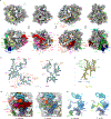
. 2019 Nov;4(11):1798-1804.
doi: 10.1038/s41564-019-0514-6. Epub 2019 Jul 22.

Evolutionary compaction and adaptation visualized by the structure of the dormant microsporidian ribosome

Affiliations

Evolutionary compaction and adaptation visualized by the structure of the dormant microsporidian ribosome

Jonas Barandun et al. Nat Microbiol. 2019 Nov.

Abstract

Microsporidia are eukaryotic parasites that infect essentially all animal species, including many of agricultural importance1-3, and are significant opportunistic parasites of humans4. They are characterized by having a specialized infection apparatus, an obligate intracellular lifestyle5, rudimentary mitochondria and the smallest known eukaryotic genomes5-7. Extreme genome compaction led to minimal gene sizes affecting even conserved ancient complexes such as the ribosome8-10. In the present study, the cryo-electron microscopy structure of the ribosome from the microsporidium Vairimorpha necatrix is presented, which illustrates how genome compaction has resulted in the smallest known eukaryotic cytoplasmic ribosome. Selection pressure led to the loss of two ribosomal proteins and removal of essentially all eukaryote-specific ribosomal RNA (rRNA) expansion segments, reducing the rRNA to a functionally conserved core. The structure highlights how one microsporidia-specific and several repurposed existing ribosomal proteins compensate for the extensive rRNA reduction. The microsporidian ribosome is kept in an inactive state by two previously uncharacterized dormancy factors that specifically target the functionally important E-site, P-site and polypeptide exit tunnel. The present study illustrates the distinct effects of evolutionary pressure on RNA and protein-coding genes, provides a mechanism for ribosome inhibition and can serve as a structural basis for the development of inhibitors against microsporidian parasites.


Conflict of interest statement

Competing interests

The authors declare no competing interests.

Figures

Fig. 1 |
Fig. 1 |. Cryo-EM structure of the microsporidian ribosome reveals a prokaryotic-like rRNA core covered with eukaryotic ribosomal proteins.
a, Composite cryo-EM map consisting of the 3.26Å LSU-, the 3.3Å SSU body and the 3.64Å SSU head-focused map with rRNAs coloured in light blue (LSU) and yellow (SSU) and ribosomal proteins in shades of blue and green (LSU) or yellow and orange (SSU). Locations of msL1 (green), MDF1 (dark pink) and MDF2 (light pink) are indicated, b, Structure of the microsporidian ribosome coloured as in a with all ribosomal proteins labelled, c, Structural comparison of the E. coli (PDB 4YBB), V. necatrix, S. cerevisiae (PDB 4V88) and Homo sapiens (PDB 6EK0) ribosomes reveals the extent of microsporidian rRNA reduction. In the top panel, ribosomal proteins are shown in grey. In the middle panel, rRNAs (transparent) are superimposed on the V. necatrix rRNAs (full colours) and, in the lowest panel, rRNAs are shown as a linear schematic, nt, nucleotides.
Fig. 2 |
Fig. 2 |. Extensive rRNA expansion segment loss in microsporidia.
a, Four 90°-related views of the V. necatrix (top) and S. cerevisiae (bottom) ribosomes with LSU rRNAs coloured in light blue and SSU rRNA in pale yellow. Locations of expansion segments that have been lost in V. necatrix (top) but are present in S. cerevisiae (bottom) are coloured (SSU, shades of blue and green; LSU, shades of red and purple). b,c, Secondary structure diagram of the LSU (b) and the SSU (c) rRNAs. Lost expansion segments are coloured as in a. d, Comparison between the S. cerevisiae and the V. necatrix region around the LSU rRNA fusion site with lost expansion segments and selected ribosomal proteins coloured as in a and labelled. The msL1 is shown in light green, e, Same view as in d with selected proteins in isolation.
Fig. 3 |
Fig. 3 |. Dormancy factors blocking the E-site, the peptidyl-transf erase centre and the polypeptide exit tunnel.
a, Location of the peptidyl-transferase centre-blocking factor (MDF2, light pink) in the large ribosomal subunit and the E-site factor (MDF1, dark pink) in the small ribosomal subunit. Superimposed tRNAs (white, from PDB 4V6F) are shown in the A-, P- and E-site, b, Slab view and a zoom-in thereof showing the position of MDF1 in the E-site and MDF2 in the peptidyl-transferase centre, c, Binding of tRNAs in the P- and E-site is sterically hindered by the presence of MDF2 and MDF1, as indicated by the superimposed tRNAs shown as a transparent white surface. The location of a superimposed mRNA(grey) and the polypeptide exit tunnel (blue) is indicated.
Fig. 4 |
Fig. 4 |. MDF1 is a conserved eukaryotic protein.
a, Detailed view of the E-site blocking factor MDF1 (pink). The N-terminus of eS25 (yellow) interacts with MDF1. The inlet shows the cryo-EM density of eS25 and MDF1. b, Superposition of V. necatrix MDF1 (red) with the structure of the H sapiens (PDB 5ZRT, green) and P. falciparum (PDB 1ZSO, light blue) homologues, c, Schematic secondary structure diagram of MDF1 with the main interactions indicated, d, Sequence alignment of MDF1 and eukaryotic homologues, with secondary structure elements indicated above. The following organisms in d are not otherwise mentioned: Drosophila pseudoobscura, Drosophila melanogaster, Caenorhabditis elegans, Schizosaccharomyces pombe, Dictyostelium discoideum, Nernatostella vectensis, Rattus norvegicus, Bos taurus, Mus musculus and Xenopus laevis.

References

    1. Stentiford GD et al. Microsporidia—emergent pathogens in the global food chain. Trends Parasitol. 32, 336–348 (2016). - PMC - PubMed
    1. Franzen C Microsporidia: a review of 150 years of research. Open Parasitol. J 2, 1–34 (2008).
    1. Higes M et al. How natural infection by Nosema ceranae causes honeybee colony collapse. Environ. Microbiol 10, 2659–2669 (2008). - PubMed
    1. Didier ES & Weiss LM Microsporidiosis: not just in AIDS patients. Curr. Opin. Infect. Dis 24, 490–495 (2011). - PMC - PubMed
    1. Corradi N Microsporidia: eukaryotic intracellular parasites shaped by gene loss and horizontal gene transfers. Annu. Rev. Microbiol 69, 167–183 (2015). - PubMed

Publication types