

Twenty-four hour ocular and systemic diurnal rhythms in children
- PMID: 31332822
- PMCID: PMC7092716
- DOI: 10.1111/opo.12633
Twenty-four hour ocular and systemic diurnal rhythms in children
Abstract
Purpose: Ocular diurnal rhythms have been implicated in myopia, glaucoma, diabetes, and other ocular pathologies. Ocular rhythms have been well described in adults; however, they have not yet been fully examined in children. The goal of this study was to investigate ocular and systemic diurnal rhythms over 24 h in children.
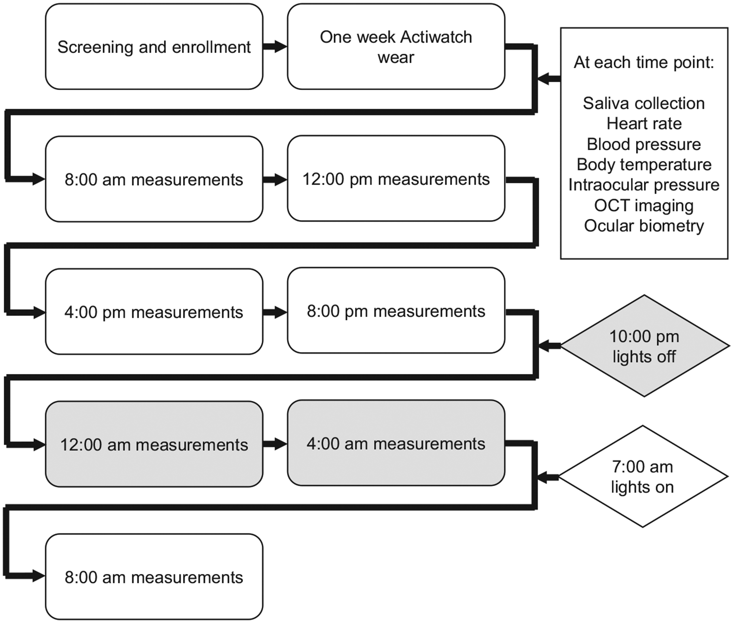
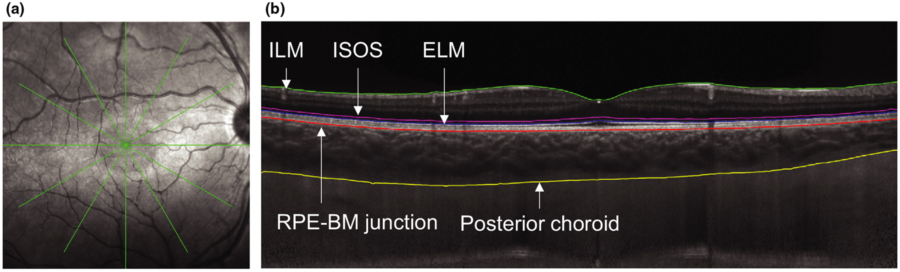
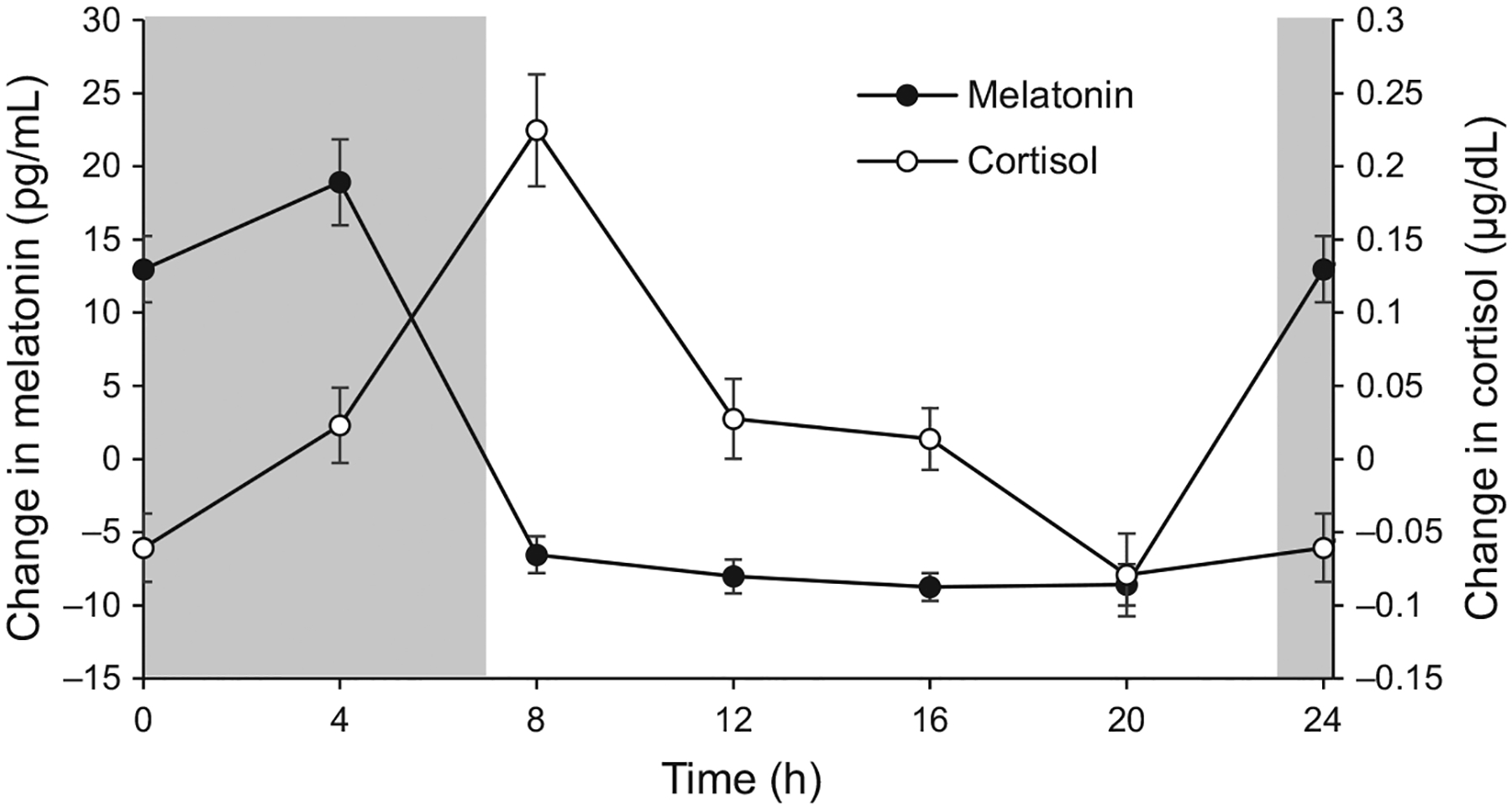
Methods: Subjects, ages 5 to 14 years (n = 18), wore a light, sleep, and activity monitor for one week to assess habitual sleep/wake patterns, then underwent diurnal measurements every 4 h for 24 h. Measurements included blood pressure, heart rate, body temperature, intraocular pressure (IOP), ocular biometry, and optical coherence tomography imaging. Saliva was collected for melatonin and cortisol analysis. Mean ocular perfusion pressure was calculated from IOP and blood pressure. Central corneal thickness, corneal power, anterior chamber depth, lens thickness, vitreous chamber depth, and axial length were determined from biometry. Total retinal thickness, retinal pigment epithelium (RPE) + photoreceptor outer segment thickness, photoreceptor inner segment thickness, and choroidal thickness were determined for a 1 mm diameter centred on the fovea. Subjects' amplitude and acrophase of diurnal variation for each parameter were determined using Fourier analysis, and mean acrophase was calculated using unit vector averaging.
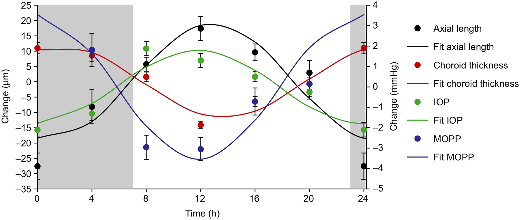
Results: Repeated measures analysis of variance (ANOVA) showed that all parameters except anterior chamber depth exhibited significant variations over 24 h (p ≤ 0.005 for all). Axial length underwent diurnal variation of 45.25 ± 6.30 μm with an acrophase at 12.92 h, and choroidal thickness underwent diurnal variation of 26.25 ± 2.67 μm with an acrophase at 1.90 h. IOP was approximately in phase with axial length, with a diurnal variation of 4.19 ± 0.50 mmHg and acrophase at 11.37 h. Total retinal thickness underwent a significant diurnal variation of 4.09 ± 0.39 μm with an acrophase at 15.04 h. The RPE + outer segment layer was thickest at 3.25 h, while the inner segment layer was thickest at 14.95 h. Melatonin peaked during the dark period at 2.36 h, and cortisol peaked after light onset at 9.22 h.
Conclusions: Ocular and systemic diurnal rhythms were robust in children and similar to those previously reported in adult populations. Axial length and IOP were approximately in phase with each other, and in antiphase to choroidal thickness. These findings may have important implications in myopia development in children.
Keywords: axial length; choroidal thickness; circadian rhythms; cortisol; diurnal rhythms; melatonin.
© 2019 The Authors Ophthalmic & Physiological Optics © 2019 The College of Optometrists.
Conflict of interest statement
Conflict of interest
The authors report no conflicts of interest and have no proprietary interest in any of the materials mentioned in this article.
Figures

References
-
- Aschoff J Circadian rhythms: influences of internal and external factors on the period measured in constant conditions. Z Tierpsychol 1979; 49: 225–249. - PubMed
-
- Dacey DM, Liao HW, Peterson BB et al. Melanopsin-expressing ganglion cells in primate retina signal colour and irradiance and project to the LGN. Nature 2005; 433: 749–754. - PubMed
-
- Gillette MU & Tischkau SA. Suprachiasmatic nucleus: the brain’s circadian clock. Recent Prog Horm Res 1999; 54: 33–58. - PubMed
-
- Spaide RF, Koizumi H & Pozzoni MC. Enhanced depth imaging spectral-domain optical coherence tomography. Am J Ophthalmol 2008; 146: 496–500. - PubMed
Publication types
MeSH terms
Grants and funding
LinkOut - more resources
Full Text Sources
