Review
. 2019 Jul 5:13:689.
doi: 10.3389/fnins.2019.00689. eCollection 2019.

Tissue Response to Neural Implants: The Use of Model Systems Toward New Design Solutions of Implantable Microelectrodes

Affiliations
Review

Tissue Response to Neural Implants: The Use of Model Systems Toward New Design Solutions of Implantable Microelectrodes

Maurizio Gulino et al. Front Neurosci. .

Abstract

The development of implantable neuroelectrodes is advancing rapidly as these tools are becoming increasingly ubiquitous in clinical practice, especially for the treatment of traumatic and neurodegenerative disorders. Electrodes have been exploited in a wide number of neural interface devices, such as deep brain stimulation, which is one of the most successful therapies with proven efficacy in the treatment of diseases like Parkinson or epilepsy. However, one of the main caveats related to the clinical application of electrodes is the nervous tissue response at the injury site, characterized by a cascade of inflammatory events, which culminate in chronic inflammation, and, in turn, result in the failure of the implant over extended periods of time. To overcome current limitations of the most widespread macroelectrode based systems, new design strategies and the development of innovative materials with superior biocompatibility characteristics are currently being investigated. This review describes the current state of the art of in vitro, ex vivo, and in vivo models available for the study of neural tissue response to implantable microelectrodes. We particularly highlight new models with increased complexity that closely mimic in vivo scenarios and that can serve as promising alternatives to animal studies for investigation of microelectrodes in neural tissues. Additionally, we also express our view on the impact of the progress in the field of neural tissue engineering on neural implant research.

Keywords: brain slice cultures; deep brain stimulation; foreign body reaction; microelectrodes; neural tissue engineering; neural tissue response.


Figures

FIGURE 1
FIGURE 1
Schematic timeline representation of the reactions involved in the process of neural tissue response to implantable microelectrodes. The acute phase of inflammation is characterized by BBB disruption and neuronal death due to mechanical insult followed by glial activation and immune cell recruitment at the injury site. Microelectrode performance may be hampered at this level due to mechanical mismatch with the tissue accompanied by a temporary recovery. In the chronic phase of inflammation, a glial fibrotic scar surrounds the microelectrode impeding material and stimulating site integrity that, ultimately, may result in implant failure.
FIGURE 2
FIGURE 2
Schematic representation of the current and promising in vitro/ex vivo models with increased physiological relevance for the screening of materials and coatings for the development of implantable microelectrodes.
FIGURE 3
FIGURE 3
Schematic representations of 3D microfluidic systems. (A) Schematic view of a microfluidic device for 3D cell culture composed by a vascular channel (VC) for primary human brain-derived microvascular endothelial cells (hBMVEC), and a brain chamber for primary cell-derived human neurons, pericytes and astrocytes culture in a type I collagen matrix. Reprinted from Brown et al. (2015) with the permission of AIF publishing. (B) 3D microfluidic platform for the establishment of a neurovascular unit (NVU) including blood-brain barrier (BBB). The NVU is characterized by a VC composed by a co-culture of HUVEC and Primary human lung fibroblasts, and a secondary NC composed by a co-culture of neurons and astrocytes. Adapted with permission from Bang et al. (2017). (C) Organ-on-a-chip device for 3D culture and differentiation of brain organoids, showing an enlarged view of the component parts and a flow chart showing the development stages of hiPSCs-derived brain organoids (Wang Y. et al., 2018) published by the Royal Society of Chemistry. (D) Vertical cross-section view of a perforating multi-electrode array (MEA) integrated in a PDMS device for long-term culture, live imaging, recording and stimulation of brain tissues and 3D cultures (Killian et al., 2016).
FIGURE 4
FIGURE 4
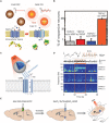
New strategies for deep brain stimulation using functional nanoparticles. (A) A schematic description of magnetothermal effect on transient receptor potential cation channel subfamily V member 1 (TRPV1) cells. (B) Comparison of the neuron reactivity under different conditions. Figures from Chen et al. (2015), reprinted with permission from AAAS. (C) A schematic description of nanoparticle-mediated near infrared (NIR) upconversion optogenetics. (D) Hippocampal local field potential response under NIR stimulation under different conditions. (E) In vivo experimental description of NIR stimulation of the ventral tegmental area of mice. Figures from Chen S. et al. (2018), reprinted with permission from AAAS.

References

    1. Achyuta A. K. H., Polikov V. S., White A. J., Pryce Lewis H. G., Murthy S. K. (2010). Biocompatibility assessment of insulating silicone polymer coatings using an in vitro glial scar assay. Macromol. Biosci. 10 872–880. 10.1002/mabi.200900451 - DOI - PubMed
    1. Adjei I., Sharma B., Labhasetwar V. (2014). Nanoparticles: cellular uptake and cytotoxicity. Adv. Exp. Med. Biol. 811 73–91. 10.1007/978-94-017-8739-0_5 - DOI - PubMed
    1. Adriani G., Ma D., Pavesi A., Kamm R. D., Goh E. L. K. (2017). A 3D neurovascular microfluidic model consisting of neurons, astrocytes and cerebral endothelial cells as a blood–brain barrier. Lab Chip 17 448–459. 10.1039/c6lc00638h - DOI - PubMed
    1. Allitt B. J., Harris A. R., Morgan S. J., Clark G. M., Paolini A. G. (2016). Thin-film micro-electrode stimulation of the cochlea in rats exposed to aminoglycoside induced hearing loss. Hear. Res. 331 13–26. 10.1016/j.heares.2015.10.003 - DOI - PubMed
    1. Antensteiner M., Abidian M. R. (2017). Tunable nanostructured conducting polymers for neural interface applications. Conf. Proc. IEEE Eng. Med. Biol. Soc. 2017 1881–1884. 10.1109/EMBC.2017.8037214.Tunable - DOI - PMC - PubMed

LinkOut - more resources