Review
. 2019 Jul 5:10:751.
doi: 10.3389/fphar.2019.00751. eCollection 2019.

Bioinspired and Biomimetic Nanotherapies for the Treatment of Infectious Diseases

Affiliations
Review

Bioinspired and Biomimetic Nanotherapies for the Treatment of Infectious Diseases

Guoyu Yang et al. Front Pharmacol. .

Abstract

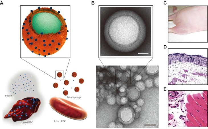
There are still great challenges for the effective treatment of infectious diseases, although considerable achievement has been made by using antiviral and antimicrobial agents varying from small-molecule drugs, peptides/proteins, to nucleic acids. The nanomedicine approach is emerging as a new strategy capable of overcoming disadvantages of molecular therapeutics and amplifying their anti-infective activities, by localized delivery to infection sites, reducing off-target effects, and/or attenuating resistance development. Nanotechnology, in combination with bioinspired and biomimetic approaches, affords additional functions to nanoparticles derived from synthetic materials. Herein, we aim to provide a state-of-the-art review on recent progress in biomimetic and bioengineered nanotherapies for the treatment of infectious disease. Different biomimetic nanoparticles, derived from viruses, bacteria, and mammalian cells, are first described, with respect to their construction and biophysicochemical properties. Then, the applications of diverse biomimetic nanoparticles in anti-infective therapy are introduced, either by their intrinsic activity or by loading and site-specifically delivering various molecular drugs. Bioinspired and biomimetic nanovaccines for prevention and/or therapy of infectious diseases are also highlighted. At the end, major translation issues and future directions of this field are discussed.

Keywords: bioengineering; biomimetic; infectious disease; nanoparticle; targeted therapy; vaccine.


Figures

Figure 1
Figure 1
Schematic illustration of virus-mimetic nanoparticles. (A) Virosomes. (B) Virus-like particles (VLPs). (C) Self-assembling nanoparticles displaying antigens on their surface. (D) Fully synthetic nanoparticles mimicking viruses.
Figure 2
Figure 2
Molecular design and characterization of virus-mimicking self-assembling nanoparticles. (A, B) Construction of hemagglutinin (HA)-ferritin fusion protein-assembled nanoparticles displaying influenza virus HA. (A) A subunit of H. pylori non-haem ferritin [protein data bank (PDB: 3bve)] (left). The NH2- and COOH-termini are labeled as N and C, respectively. Three subunits surrounding a threefold axis are shown (middle) and Asp 5 is colored in red. An assembled ferritin nanoparticle and an HA trimer (PDB: 3sm5) (viewed from membrane proximal end) (right). A triangle connecting the Asp 5 residues at the threefold axis is shown in red. The same triangle is drawn on the HA trimer (right). A schematic representation of the HA-ferritin fusion protein is shown (bottom). (B) Negatively stained transmission electron microscopy (TEM) images of HA nanoparticles. (C–E) Design, in vitro assembly, and structural characterization of DS-Cav1-I53-50. (C) Schematic representation of the computational docking protocol used to identify nanoparticle components suitable for fusion to DS-Cav1. The C termini of the foldon and N termini of the nanoparticle trimer are shown as red and blue spheres, respectively, and the exterior and interior surfaces of the nanoparticle are depicted. (D) Structural model of DS-Cav1-I53-50 and schematic of the in vitro assembly process. Each nanoparticle comprises 20 trimeric and 12 pentameric building blocks for a total of 60 copies of each subunit. (E) Negatively stained TEM image of I53-50 and DS-Cav1-I53-50 nanoparticles. The two images on the right are averages of negatively stained particles. Images (A) and (B) are reprinted with permission from Kanekiyo et al. (2013). © (2013) Macmillan Publishers Limited. Images (C)–(E) are reprinted with permission from Marcandalli et al. (2019). © (2019) Elsevier Inc.
Figure 3
Figure 3
Engineering of cell-mimetic nanoparticles. (A) Schematic illustration of the preparation process of the red blood cell (RBC) membrane-coated poly(lactic-co-glycolic acid) (PLGA) nanoparticles. (B) Representative TEM images of uranyl acetate-stained RBC membrane-coated PLGA nanoparticles (NPs). (C–F) Synthesis, formulation, and characterization of leukosomes. (C) Extraction of proteolipid materials from murine J774 macrophages. (D) Protein enrichment of the phospholipid film. (E) Vesicular formulation of leukosomes. (F) Cryo-TEM analysis of leukosomes. NPs, nanoparticles; DPPC, 1,2-dihexadecanoyl-sn-glycero-3-phosphocholine; DSPC, 1,2-distearoyl-sn-glycero-3-phosphocholine; DOPC, 1, 2-dioleoyl-sn-glycero-3-phosphocholine; CHOL, cholesterol. Images (A) and (B) are reprinted with permission from Hu et al. (2011). © (2011) National Academy of Sciences. Images (C)–(F) are reprinted with permission from Molinaro et al. (2016). © (2016) Macmillan Publishers Limited.
Figure 4
Figure 4
Design of biomimetic antiviral nanotherapies. (A) Nested symmetrical assembly of virus-like glycodendrinanoparticles using a tag-and-modify strategy. Glycodendrons are created through iterative multivalent assembly and then attached to multiple tags, each in a monomer protein. (B) Cartoon of the virucidal activity of broad-spectrum antiviral nanoparticles MUS : OT-NPs compared to MES-NPs. HSPG, heparin sulfate proteoglycans; MES, 3-mercaptoethylsulfonate; MUS, undecanesulfonic acid; OT, 1-octanethiol. Image (A) is reprinted with permission from Ribeiro-Viana et al. (2012). © (2012) Macmillan Publishers Limited. Image (B) is reprinted with permission from Cagno et al. (2018). © (2018) Macmillan Publishers Limited.
Figure 5
Figure 5
Schematic, characterization, and in vivo evaluation of a pore-forming toxin-absorbing biomimetic nanosponge. (A) Schematic of a toxin nanosponge and the mechanism of neutralizing pore-forming toxins. The nanosponge consists of substrate-supported RBC bilayer membranes into which pore-forming toxins can be incorporated. (B) TEM images of nanosponges mixed with α-toxin (scale bar, 80 nm) and the zoomed-in view of a single toxin-absorbed nanosponge (scale bar, 20 nm). (C–E) Mice injected with α -toxin/nanosponges. No skin lesion occurred (C). No abnormality was observed in the epidermis (D). Normal muscle structure was observed (E). Images are reprinted with permission from (Hu et al., 2013a). © (2013) Macmillan Publishers Limited.

Similar articles

Cited by

References

    1. Abdoli A., Soleimanjahi H., Tavassoti Kheiri M., Jamali A., Mazaheri V., Abdollahpour Alitappeh M. (2014). An h1-h3 chimeric influenza virosome confers complete protection against lethal challenge with pr8 (h1n1) and x47 (h3n2) viruses in mice. Pathog. Dis. 72 (3), 197–207. 10.1111/2049-632X.12206 - DOI - PubMed
    1. Abed N., Couvreur P. (2014). Nanocarriers for antibiotics: a promising solution to treat intracellular bacterial infections. Int. J. Antimicrob. Agents 43 (6), 485–496. 10.1016/j.ijantimicag.2014.02.009 - DOI - PubMed
    1. Ahmadi Badi S., Moshiri A., Fateh A., Rahimi Jamnani F., Sarshar M., Vaziri F., et al. (2017). Microbiota-derived extracellular vesicles as new systemic regulators. Front. Microbiol. 8, 1610–1610. 10.3389/fmicb.2017.01610 - DOI - PMC - PubMed
    1. Al-Halifa S., Gauthier L., Arpin D., Bourgault S., Archambault D. (2019). Nanoparticle-based vaccines against respiratory viruses. Front. Immunol. 10, 11. 10.3389/fimmu.2019.00022 - DOI - PMC - PubMed
    1. Almeida J., Edwards D. C., Brand C., Heath T. (1975). Formation of virosomes from influenza subunits and liposomes. Lancet 306 (7941), 899–901. 10.1016/S0140-6736(75)92130-3 - DOI - PubMed

LinkOut - more resources