. 2019 Jul 5:10:863.
doi: 10.3389/fphys.2019.00863. eCollection 2019.

Identification of a PEST Sequence in Vertebrate KIR2.1 That Modifies Rectification

Affiliations

Identification of a PEST Sequence in Vertebrate KIR2.1 That Modifies Rectification

Muge Qile et al. Front Physiol. .

Abstract

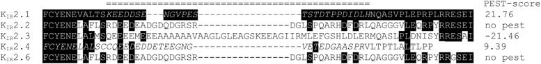
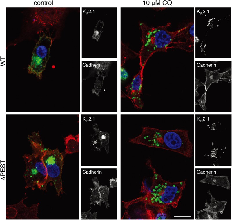
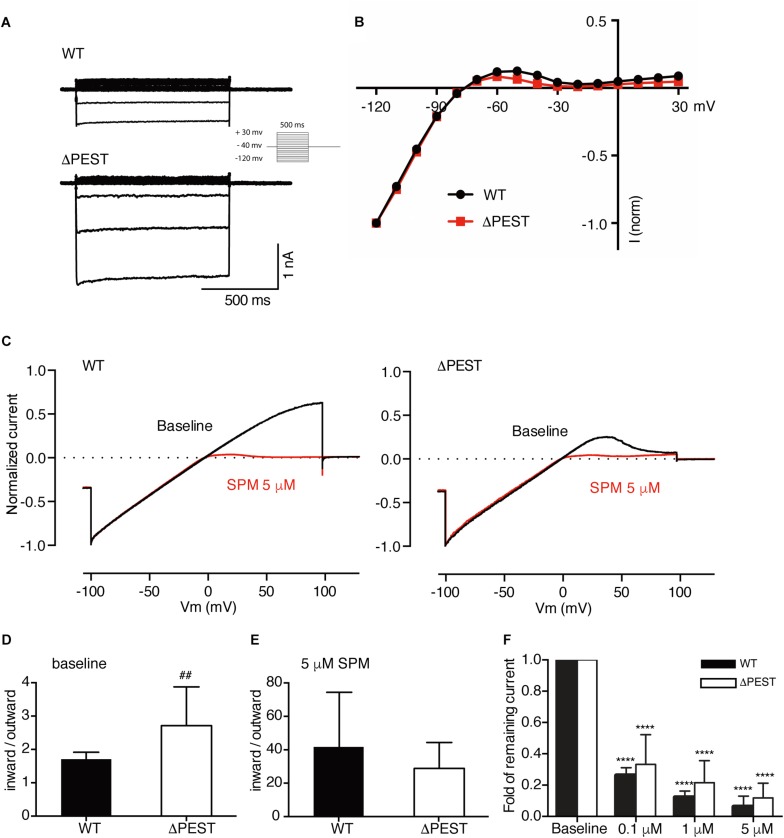
KIR2.1 potassium channels, producing inward rectifier potassium current (I K1 ), are important for final action potential repolarization and a stable resting membrane potential in excitable cells like cardiomyocytes. Abnormal KIR2.1 function, either decreased or increased, associates with diseases such as Andersen-Tawil syndrome, long and short QT syndromes. KIR2.1 ion channel protein trafficking and subcellular anchoring depends on intrinsic specific short amino acid sequences. We hypothesized that combining an evolutionary based sequence comparison and bioinformatics will identify new functional domains within the C-terminus of the KIR2.1 protein, which function could be determined by mutation analysis. We determined PEST domain signatures, rich in proline (P), glutamic acid (E), serine (S), and threonine (T), within KIR2.1 sequences using the "epestfind" webtool. WT and ΔPEST KIR2.1 channels were expressed in HEK293T and COS-7 cells. Patch-clamp electrophysiology measurements were performed in the inside-out mode on excised membrane patches and the whole cell mode using AxonPatch 200B amplifiers. KIR2.1 protein expression levels were determined by western blot analysis. Immunofluorescence microscopy was used to determine KIR2.1 subcellular localization. An evolutionary conserved PEST domain was identified in the C-terminus of the KIR2.1 channel protein displaying positive PEST scores in vertebrates ranging from fish to human. No similar PEST domain was detected in KIR2.2, KIR2.3, and KIR2.6 proteins. Deletion of the PEST domain in California kingsnake and human KIR2.1 proteins (ΔPEST), did not affect plasma membrane localization. Co-expression of WT and ΔPEST KIR2.1 proteins resulted in heterotetrameric channel formation. Deletion of the PEST domain did not increase protein stability in cycloheximide assays [T½ from 2.64 h (WT) to 1.67 h (ΔPEST), n.s.]. WT and ΔPEST channels, either from human or snake, produced typical I K1 , however, human ΔPEST channels displayed stronger intrinsic rectification. The current observations suggest that the PEST sequence of KIR2.1 is not associated with rapid protein degradation, and has a role in the rectification behavior of I K1 channels.

Keywords: KIR2.1; PEST domain; channel; inward rectifier; patch clamp; potassium; vertebrates.


Figures

FIGURE 1
FIGURE 1
Amino acid alignment of C-termini of human KIR2.1, KIR2.2, KIR2.3, KIR2.4, and KIR2.6 encompassing the PEST domain region of KIR2.1 indicated by double line above the alignment. Amino acid sequences are depicted in single letter code. Identical residues with respect to KIR2.1 are depicted in white font on a black background. KIR2.4 contains a potential PEST sequence extending from 378 to 424 (KSSFPGSLTAFCYENELALSCCQEEDEDDETEEGNGVETEDGAASPR). PEST domains in KIR2.1 and KIR2.4 are indicated in italic. PEST scores are depicted at the right side of the sequences.
FIGURE 2
FIGURE 2
Expression analysis and channel formation of human WT and ΔPEST KIR2.1 protein. (A) Western blot depicting WT (approximately 50 kDa) and ΔPEST (approximately 47 kDa) KIR2.1 proteins expressed in HEK293T cells. Non-transfected cells (NT) were used as negative control. Ponceau staining depicts loading control. (B) Subcellular localization of ectopically expressed WT and ΔPEST KIR2.1 channel proteins in COS-7 cells. Arrows indicate membrane ruffles with KIR2.1 expression. (C) HEK293T cells were co-transfected with GFP-tagged murine KIR2.1 and either WT or ΔPEST KIR2.1. Non-transfected cells (NT) were used as negative control. KIR2.1-GFP was detected by GFP antibody (WB: GFP) for IP control, and N-terminal KIR2.1 antibody (WB: KIR2.1) was used to detect KIR2.1-GFP either WT or ΔPEST non-tagged KIR2.1 protein. Positions of KIR2.1-GFP, WT-KIR2.1 and ΔPEST-KIR2.1 are indicated. Lysate blots serve as immune-precipitation input control. *IgG heavy chain.
FIGURE 3
FIGURE 3
CQ treatment induces intracellular accumulation of WT and ΔPEST KIR2.1 protein in COS-7 cells. Confocal images of WT and ΔPEST KIR2.1 detected by N-terminal KIR2.1 antibody (green), and Cadherin (membrane staining) by Pan-Cadherin antibody (red). Single staining results are depicted on the right by b/w images. Scale bar indicates 10 μm.
FIGURE 4
FIGURE 4
Cycloheximide (CHX) assay of KIR2.1 degradation in transfected HEK293T cells. (A) Example of WT and ΔPEST KIR2.1 protein degradation after exposure to 200 μg/mL CHX for different time intervals. Non-transfected (NT) cell were used as negative control. (B) Quantification of CHX assays to depict normalized KIR2.1 expression vs. timed CHX treatment. Dotted line indicates 50% of initial normalized KIR2.1 protein signal. ∗∗P < 0.01 WT vs. ΔPEST.
FIGURE 5
FIGURE 5
Electrophysiological analysis of human WT and ΔPEST KIR2.1 channels transiently transfected in HEK293T cells. (A) Representative current traces of WT and ΔPEST IKIR2.1 recorded in whole cell mode. (B) Normalized current–voltage relation curve of WT and ΔPEST IKIR2.1 (mean ± SEM; WT n = 18, ΔPEST n = 18), note that error bars are smaller than symbols at each point. (C) Steady state IKIR2.1 traces from WT and ΔPEST channel containing excised inside–out patches elicited by a voltage ramp protocol from -100 to + 100 mV over 5 s, under baseline conditions (black) and upon application of 5 μM spermine (red). (D,E) Quantification of rectification index (inward current at –80 mV divided by outward current at +50 mV) of WT and ΔPEST IKIR2.1 from ramp protocol elicited currents in inside-out mode without (D, baseline) and in the presence of 5 μM spermine (E) (mean ± SD, WT n = 10, ΔPEST n = 10). (F) Quantification of normalized outward current (at +50 mV) from WT and ΔPEST channels in inside-out patch clamp under baseline conditions and with increasing spermine concentrations. ##P < 0.01 vs. WT; ****P < 0.0001 vs. baseline (mean ± SD, WT n = 10, ΔPEST n = 10).
FIGURE 6
FIGURE 6
Expression analysis and channel formation of snake WT and ΔPEST KIR2.1 protein and electrophysiological analysis of formed channels in transiently transfected HEK293T cells and COS-7 cells. (A) Western blot depicting WT (approximately 50 kDa) and ΔPEST (approximately 47 kDa) KIR2.1 protein. Non-transfected cells (NT) were used as negative control. Ponceau staining depicts loading control. (B) Subcellular localization of ectopically expressed WT and ΔPEST KIR2.1 channel proteins in COS-7 cells. Apart from plasma membrane staining, intracellular aggregates were observed. (C) Representative current traces of WT and ΔPEST IKIR2.1 recorded in whole cell mode (left) and normalized current–voltage relation curves of WT and ΔPEST IKIR2.1 (right) (mean ± SEM, WT n = 9, ΔPEST n = 7). (D) Steady state IKIR2.1 traces from WT and ΔPEST channel containing inside–out patches elicited by a voltage ramp protocol from -100 to + 100 mV, under baseline conditions (black) and upon application of 5 μM spermine (red). (E) Quantification of rectification index (inward current at –80 mV divided by outward current at +50 mV) of WT and ΔPEST IKIR2.1 from ramp protocol elicited currents in inside-out mode without (left panel) and in the presence of 5 μM spermine (middle panel) (mean ± SD, WT n = 11, ΔPEST n = 24). Quantification of normalized outward current (at +50 mV) from WT and ΔPEST channels in inside-out patches under baseline conditions and with increasing spermine concentrations (right panel). *P < 0.05 vs. baseline (mean ± SD, WT n = 10, ΔPEST n = 10).

References

    1. Belizario J. E., Alves J., Garay-Malpartida M., Occhiucci J. M. (2008). Coupling caspase cleavage and proteasomal degradation of proteins carrying PEST motif. Curr. Protein Pept. Sci. 9 210–220. 10.2174/138920308784534023 - DOI - PubMed
    1. Bies J., Markus J., Wolff L. (2002). Covalent attachment of the SUMO-1 protein to the negative regulatory domain of the c-Myb transcription factor modifies its stability and transactivation capacity. J. Biol. Chem. 277 8999–9009. 10.1074/jbc.M110453200 - DOI - PubMed
    1. De Boer T. P., Houtman M. J., Compier M., Van der Heyden M. A. G. (2010). The mammalian KIR2.x inward rectifier ion channel family: expression pattern and pathophysiology. Acta Physiol. 199 243–256. 10.1111/j.1748-1716.2010.02108.x - DOI - PubMed
    1. Gokhale R. S., Khosla C. (2000). Role of linkers in communication between protein modules. Curr. Opin. Chem. Biol. 4 22–27. 10.1016/S1367-5931(99)00046-0 - DOI - PubMed
    1. Hansen S. B., Tao X., MacKinnon R. (2011). Structural basis of PIP2 activation of the classical inward rectifier K+ channel Kir2.2. Nature 477 495–498. 10.1038/nature10370 - DOI - PMC - PubMed