

Dapagliflozin Attenuates Renal Tubulointerstitial Fibrosis Associated With Type 1 Diabetes by Regulating STAT1/TGFβ1 Signaling
- PMID: 31333586
- PMCID: PMC6616082
- DOI: 10.3389/fendo.2019.00441
Dapagliflozin Attenuates Renal Tubulointerstitial Fibrosis Associated With Type 1 Diabetes by Regulating STAT1/TGFβ1 Signaling
Abstract
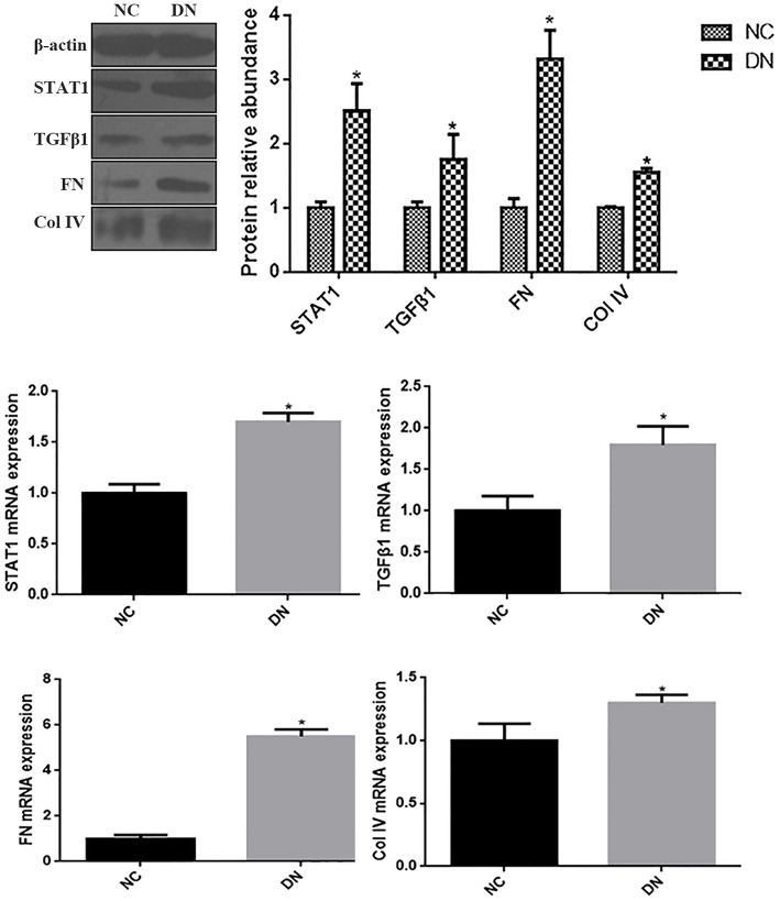
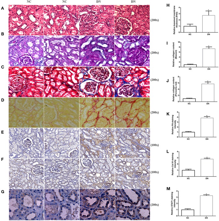
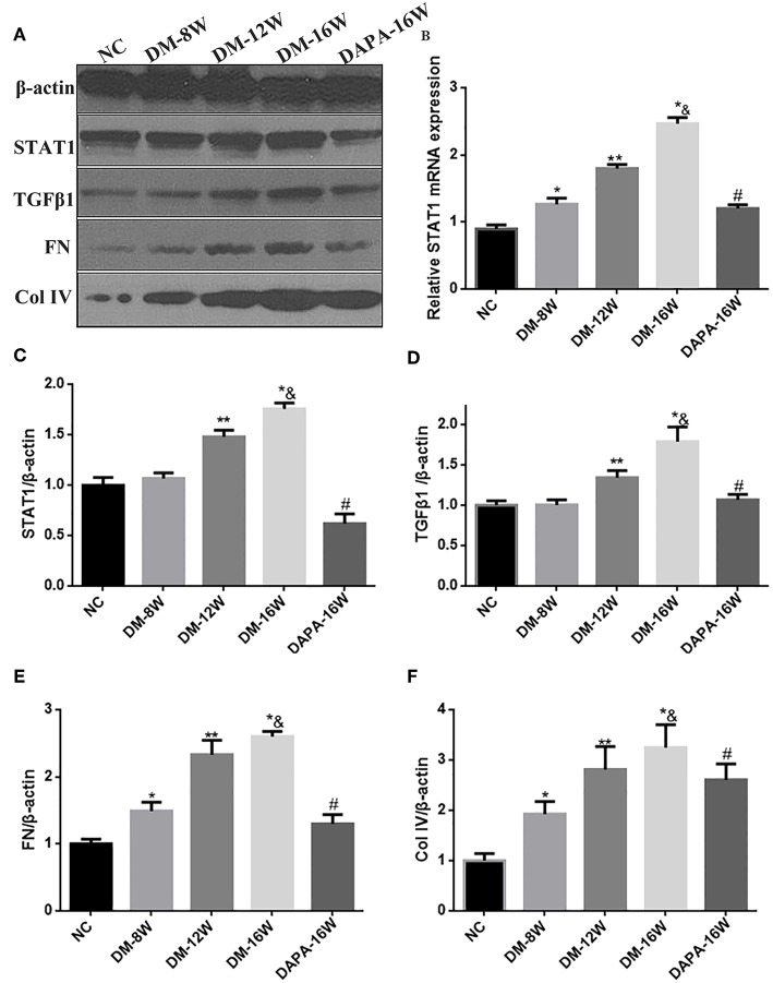
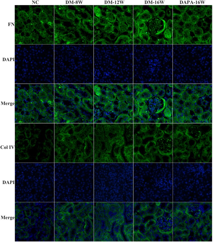
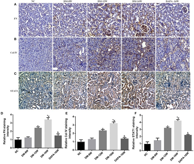
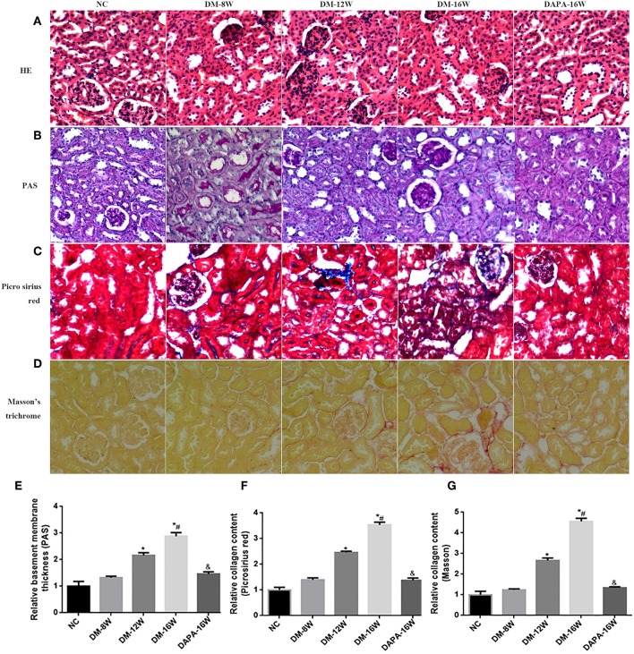
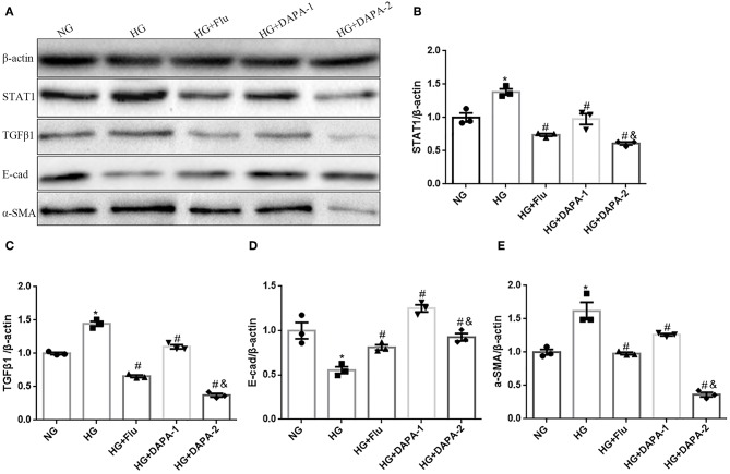
Tubulointerstitial fibrosis (TIF) plays an important role in the progression of renal fibrosis in diabetic nephropathy (DN). Accumulating evidence supports a crucial inhibitory effect of dapagliflozin, a SGLT2 inhibitor, on TIF, but the underlying mechanisms remain largely unknown. This study aimed to shed light on the efficacy of dapagliflozin in reducing TIF as well as its possible impact on renal function. TIF in human kidney biopsies obtained from patients with DN was quantified by histopathological staining. In vitro, HK-2 cells were incubated in high glucose with dapagliflozin or fludarabine, and epithelial-mesenchymal transition (EMT) was determined. In vivo experiments were performed in streptozotocin (STZ)-induced type 1 diabetic mice treated with dapagliflozin by gavage for 16 weeks, after which specific functional characteristics and TIF were analyzed. In both DN patients and diabetic mice, fibronectin and Col IV, as well as STAT1 protein in the kidneys were increased as compared with controls. Dapagliflozin significantly decreased blood glucose, and renal STAT1 and TGF-β1 expression in mice. Furthermore, dapagliflozin improved renal function, and attenuated diabetes-induced TIF. In HK-2 cells, dapagliflozin, and fludarabine directly decreased aberrant STAT1 expression and reversed high glucose-induced downregulation of E-cadherin and α-SMA induction. Thus, the results demonstrate that dapagliflozin not only improves hyperglycemia but also slows down the progression of diabetes-associated renal TIF by improving hyperglycemia-induced activation of the STAT1/TGF-β1 pathway.
Keywords: STAT1; TGF-β1; dapagliflozin; epithelial mesenchymal transition; tubulointerstitial fibrosis.
Figures
Similar articles
-
FSP1-specific SMAD2 knockout in renal tubular, endothelial, and interstitial cells reduces fibrosis and epithelial-to-mesenchymal transition in murine STZ-induced diabetic nephropathy.Cell Tissue Res. 2018 Apr;372(1):115-133. doi: 10.1007/s00441-017-2754-1. Epub 2017 Dec 6. Cell Tissue Res. 2018. PMID: 29209813
-
FoxO1-mediated inhibition of STAT1 alleviates tubulointerstitial fibrosis and tubule apoptosis in diabetic kidney disease.EBioMedicine. 2019 Oct;48:491-504. doi: 10.1016/j.ebiom.2019.09.002. Epub 2019 Oct 16. EBioMedicine. 2019. PMID: 31629675 Free PMC article.
-
Jujuboside A ameliorates tubulointerstitial fibrosis in diabetic mice through down-regulating the YY1/TGF-β1 signaling pathway.Chin J Nat Med. 2022 Sep;20(9):656-668. doi: 10.1016/S1875-5364(22)60200-0. Chin J Nat Med. 2022. PMID: 36162951
-
High glucose down-regulates microRNA-181a-5p to increase pro-fibrotic gene expression by targeting early growth response factor 1 in HK-2 cells.Cell Signal. 2017 Feb;31:96-104. doi: 10.1016/j.cellsig.2017.01.012. Epub 2017 Jan 7. Cell Signal. 2017. PMID: 28077323
-
miR-133b and miR-199b knockdown attenuate TGF-β1-induced epithelial to mesenchymal transition and renal fibrosis by targeting SIRT1 in diabetic nephropathy.Eur J Pharmacol. 2018 Oct 15;837:96-104. doi: 10.1016/j.ejphar.2018.08.022. Epub 2018 Aug 17. Eur J Pharmacol. 2018. PMID: 30125566
Cited by
-
The roles of the ubiquitin-proteasome system in renal disease.Int J Med Sci. 2025 Mar 10;22(8):1791-1810. doi: 10.7150/ijms.107284. eCollection 2025. Int J Med Sci. 2025. PMID: 40225869 Free PMC article. Review.
-
Ellagic acid ameliorates renal fibrogenesis by blocking epithelial-to-mesenchymal transition.Tzu Chi Med J. 2023 Oct 24;36(1):59-66. doi: 10.4103/tcmj.tcmj_106_23. eCollection 2024 Jan-Mar. Tzu Chi Med J. 2023. PMID: 38406569 Free PMC article.
-
LncRNA MEG3 inhibits renal fibrinoid necrosis of diabetic nephropathy via the MEG3/miR-21/ORAI1 axis.Mol Biol Rep. 2023 Apr;50(4):3283-3295. doi: 10.1007/s11033-023-08254-2. Epub 2023 Jan 30. Mol Biol Rep. 2023. PMID: 36715789
-
Gentiopicroside Ameliorates Diabetic Renal Tubulointerstitial Fibrosis via Inhibiting the AT1R/CK2/NF-κB Pathway.Front Pharmacol. 2022 Jun 23;13:848915. doi: 10.3389/fphar.2022.848915. eCollection 2022. Front Pharmacol. 2022. PMID: 35814242 Free PMC article.
-
Evaluation of the novel ALK5 inhibitor EW-7197 on therapeutic efficacy in renal fibrosis using a three-dimensional chip model.Kidney Res Clin Pract. 2025 Jul;44(4):612-625. doi: 10.23876/j.krcp.23.324. Epub 2024 Sep 9. Kidney Res Clin Pract. 2025. PMID: 39384357 Free PMC article.
References
LinkOut - more resources
Full Text Sources
Research Materials
Miscellaneous