Review
. 2019 Jul 3:10:1462.
doi: 10.3389/fimmu.2019.01462. eCollection 2019.

The Metabolic Signature of Macrophage Responses

Affiliations
Review

The Metabolic Signature of Macrophage Responses

Antonella Viola et al. Front Immunol. .

Abstract

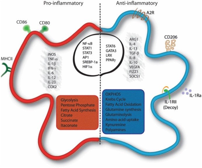
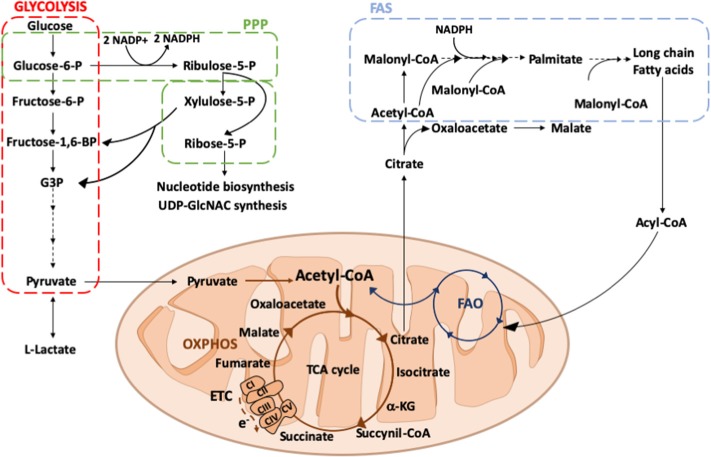
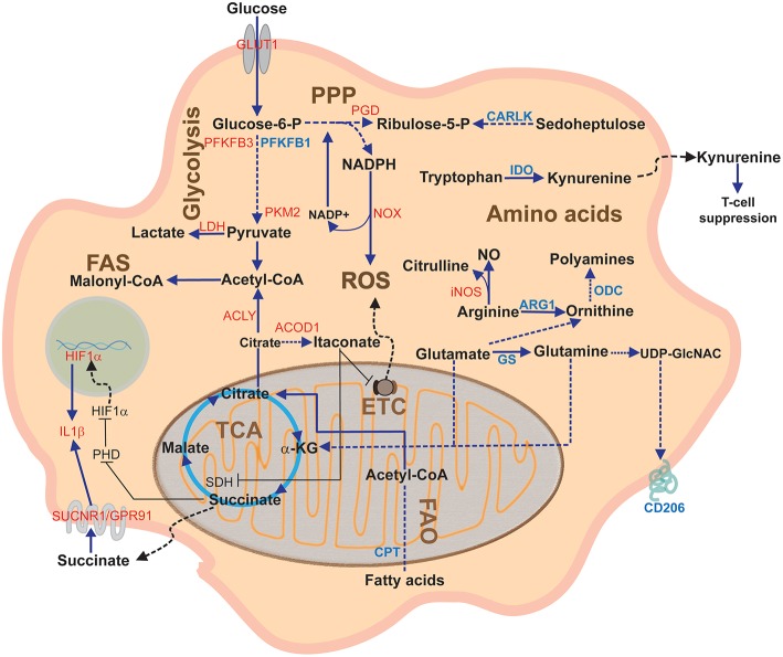
Macrophages are a heterogeneous population of immune cells playing several and diverse functions in homeostatic and immune responses. The broad spectrum of macrophage functions depends on both heterogeneity and plasticity of these cells, which are highly specialized in sensing the microenvironment and modify their properties accordingly. Although it is clear that macrophage phenotypes are difficult to categorize and should be seen as plastic and adaptable, they can be simplified into two extremes: a pro-inflammatory (M1) and an anti-inflammatory/pro-resolving (M2) profile. Based on this definition, M1 macrophages are able to start and sustain inflammatory responses, secreting pro-inflammatory cytokines, activating endothelial cells, and inducing the recruitment of other immune cells into the inflamed tissue; on the other hand, M2 macrophages promote the resolution of inflammation, phagocytose apoptotic cells, drive collagen deposition, coordinate tissue integrity, and release anti-inflammatory mediators. Dramatic switches in cell metabolism accompany these phenotypic and functional changes of macrophages. In particular, M1 macrophages rely mainly on glycolysis and present two breaks on the TCA cycle that result in accumulation of itaconate (a microbicide compound) and succinate. Excess of succinate leads to Hypoxia Inducible Factor 1α (HIF1α) stabilization that, in turn, activates the transcription of glycolytic genes, thus sustaining the glycolytic metabolism of M1 macrophages. On the contrary, M2 cells are more dependent on oxidative phosphorylation (OXPHOS), their TCA cycle is intact and provides the substrates for the complexes of the electron transport chain (ETC). Moreover, pro- and anti-inflammatory macrophages are characterized by specific pathways that regulate the metabolism of lipids and amino acids and affect their responses. All these metabolic adaptations are functional to support macrophage activities as well as to sustain their polarization in specific contexts. The aim of this review is to discuss recent findings linking macrophage functions and metabolism.

Keywords: immune cross-talk; inflammation; macrophage; metabolic rewiring; metabolism.


Figures

Figure 1
Figure 1
Molecular and metabolic signatures of macrophage activation. Pro-inflammatory stimuli induce the activation of specific pathways through the activation of transcription factors such as NF-κB, STAT1, STAT3, AP-1, SREBP-1, and HIF1α, which trigger the expression of markers like iNOS, COX-2, CD80, CD86, and MHC-II and the release of IL-1β, TNF-α, IFN-γ, IL-6, IL-12, and IL-23. Cells undergo a metabolic reprogramming toward glycolysis, the pentose-phosphate pathway, and fatty acid synthesis. This associates to interruption of the Krebs cycle, ROS formation and efflux of citrate, which supports NADPH and PGE2 synthesis, and succinate, which stabilizes HIF-1α. Itaconate is produced from citrate and displays antibacterial function. Anti-inflammatory macrophages are characterized by the expression of ARG1, FIZZ1, SOCS1, CD206, Adenosine receptor (A2R), and the decoy IL1RII and by the production of cytokines such as TGF-β, IL-10, IL-4, IL-13, IL-8, IL-1Ra, and VEGFA. Their profile is mainly controlled by the activity of the transcription factors STAT6, GATA3, PPARγ, and LRX. Metabolically, these cells display enhanced OXPHOS metabolism, fatty acid oxidation, glutaminolysis, tryptophan catabolism with release of kynurenine, and synthesis of polyamines. AP-1, Activator proten 1; ARG1, Arginase 1; COX2, cicloxygenase 2; FIZZ1, Found in inflammatory zone 1; iNOS, inducible Nitric Oxide Synthase; GATA3, GATA binding protein 3; HIF1α, Hypoxia-inducible factor 1-alpha; IFN-γ, Interferon gamma; LXR, Liver X receptor; MHC-II, major histocompatibility complex class 2; NF-κB, nuclear factor kappa-light-chain-enhancer of activated B cells; PPARγ, Peroxisome proliferator-activated receptor gamma; SOCS1, Suppressor of cytokine signaling 1; SREBP-1, Sterol regulatory element binding protein 1; STAT, Signal transducer and activator of transcription; TNF-α, Tumor necrosis factor alpha; TGF-β, transforming growth factor beta; VEGFA, Vascular endothelial growth factor A.
Figure 2
Figure 2
Overview of glucose and fatty acid metabolism. Glucose is converted into pyruvate by glycolysis (red square), in the cytosol. Among the glycolytic intermediates, glucose-6P can be diverted into PPP (green square) sustaining NADPH and ribose-5P production that, in turn, are used for fatty acid or nucleotide and UDP-GlcNAC synthesis, respectively. In hypoxic conditions, pyruvate is preferentially reduced to lactate, whereas in normoxic conditions it is decarboxylated into acetyl-CoA within the mitochondria. Here, acetyl-CoA enters into the TCA cycle, providing reducing agents to the ETC to generate energy. Citrate, an intermediate of the TCA cycle, can be exported into the cytosol where it participates in fatty acid synthesis (FAS; light blue square). Fatty acids can be oxidized via FAO (dark blue square) within the mitochondrial matrix thus generating acetyl-CoA to replenish the TCA cycle. PPP, pentose phosphate pathway; ETC, electron transport chain; TCA, tricarboxylic acid; OXPHOS, oxidative phosphorylation; FAS, fatty acid synthesis; FAO, fatty acid oxidation; Glucose 6-P, glucose 6-phosphate; Fructose 6-P, fructose 6-phosphate; Fructose-1,6-BP, fructose 1-6-biphosphate; G3P, glyceraldeyde 3-phospate; Acetyl-CoA, acetyl-Coenzyme A; α-KG, alpha-ketoglutarate; e-, electrons; CI, CII, CIII, CIV, CV, complex I, II, III, IV, V; UDP-GlcNAC, Uridine diphosphate N-acetylglucosamine.
Figure 3
Figure 3
Overview of macrophage metabolic pathways. This diagram depicts the main macrophage pathways during classical and alternative polarization and their components. In red, proteins upregulated in pro-inflammatory (M1) activation; in blue, proteins upregulated during anti-inflammatory (M2) activation. α-KG, alpha-ketoglutarate; ACLY, ATP citrate lyase; ARG1, arginase1; CARLK, carbohydrate kinase-like protein; CPT, carnitine palmitoyl transferase; ETC; Electron Transport Chain; FAO Fatty acid oxidation; FAS, Fatty acid synthesis; GS, glutamine synthetase; GLUT1, glucose transporter 1; IDH, Isocitrate dehydrogenase; IDO, indoleamine dioxygenase; iNOS, inducible nitric oxide synthase; LDH, lactate dehydrogenase; NO, nitric oxide; NOX, NADPH oxidase; ODC, ornithine decarboxylase; PGD, phosphogluconate dehydrogenase; PHD, prolyl hydroxylase; PPP, Pentose phosphate pathway; PFKFB3, phosphofructokinase fructose 2,6-biphosphatase B3; PKM2, pyruvate kinase M2; ROS, Reactive Oxygen Species; SDH, Succinate dehydrogenase; SUCNR1, succinate receptor 1; TCA, Tricarboxylic acid cycle or Krebs cycle.

References

    1. Tauber AI. Immunity: how elie metchnikoff changed the course of modern medicine by luba vikhanski. Bull Hist Med. (2017) 91:140–2. 10.1353/bhm.2017.0015 - DOI
    1. Hoeffel G, Wang Y, Greter M, See P, Teo P, Malleret B, et al. . Adult Langerhans cells derive predominantly from embryonic fetal liver monocytes with a minor contribution of yolk sac–derived macrophages. J Exp Med. (2012) 209:1167–81. 10.1084/jem.20120340 - DOI - PMC - PubMed
    1. Hashimoto D, Chow A, Noizat C, Teo P, Beasley MB, Leboeuf M, et al. . Tissue-resident macrophages self-maintain locally throughout adult life with minimal contribution from circulating monocytes. Immunity. (2013) 38:792–804. 10.1016/j.immuni.2013.04.004 - DOI - PMC - PubMed
    1. Han CZ, Ravichandran KS. Metabolic connections during apoptotic cell engulfment. Cell. (2011) 147:1442–5. 10.1016/j.cell.2011.12.006 - DOI - PMC - PubMed
    1. Fadok VA, Bratton DL, Konowal A, Freed PW, Westcott JY, Henson PM. Macrophages that have ingested apoptotic cells in vitro inhibit proinflammatory cytokine production through autocrine/paracrine mechanisms involving TGF-beta, PGE2, and PAF. J Clin Invest. (1998) 101:890–8. 10.1172/JCI1112 - DOI - PMC - PubMed

Publication types

LinkOut - more resources