. 2019 Jul 3:10:1502.
doi: 10.3389/fimmu.2019.01502. eCollection 2019.

Liver-Derived TGF-β Maintains the EomeshiTbetlo Phenotype of Liver Resident Natural Killer Cells

Affiliations

Liver-Derived TGF-β Maintains the EomeshiTbetlo Phenotype of Liver Resident Natural Killer Cells

Cathal Harmon et al. Front Immunol. .

Abstract

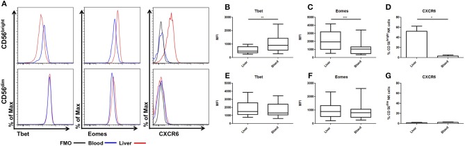
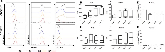
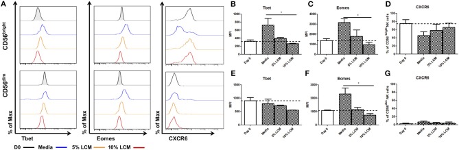
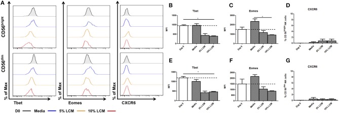
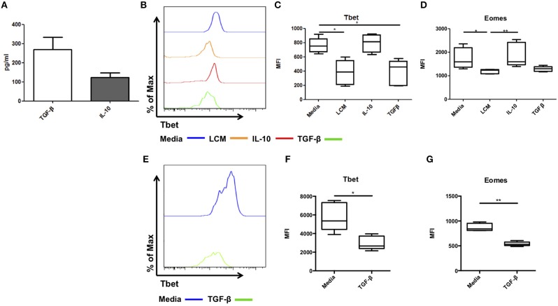
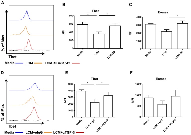
The adult human liver hosts a complex repertoire of liver resident and transient natural killer (NK) cell populations with diverse phenotypes and functions. Liver resident NK cells are CD56bright NK cells defined by a unique expression profile of transcription factors and cell surface markers (EomeshiTbetloTIGIT+CD69+CXCR6+CD49e-). Despite extensive characterization of the phenotype of liver resident NK cells, it remains unclear how factors within the liver microenvironment induce and maintain this unique phenotype. In this study, we have explored the factors regulating the phenotype of liver resident NK cells. Isolation of healthy liver resident NK cells from donor liver perfusate and in vitro culture results in the gradual loss of the characteristic Tbetlo phenotype, with the cells increasing Tbet expression significantly at day 7. This phenotypic loss could be halted through the dose-dependent addition of liver conditioned media (LCM), generated from the ex vivo culture of liver biopsies from healthy organ donors. TGF-β, but not IL-10, replicated the Tbet suppressive effects of LCM in both liver resident and peripheral blood NK cells. Furthermore, blocking TGF-β receptor signaling using the inhibitor SB431542, reversed the effect of LCM treatment on liver resident NK cells, causing the loss of tissue resident Eomeshi Tbetlo phenotype. Our findings identify liver-derived TGF-β as an important component of the liver microenvironment, which acts to regulate and maintain the phenotype of liver resident NK cells.

Keywords: Eomes; TBET; TGF-β1; liver-resident NK cell; microenviroment.


Figures

Figure 1
Figure 1
CD56bright NK cells have unique tissue resident phenotype. Mononuclear cells isolated from liver perfusate (LP) and matched peripheral blood (PB) were stained with monoclonal antibodies. NK cells were identified from the CD45+ lymphocyte gate as CD56+CD3. CD56brightCD16−/+ and CD56dimCD16+ subsets were then gated and non-viable cells were excluded by FVS780 staining. (A) Representative histograms of Tbet, Eomes and CXCR6 expression in LP and PB NK cell subsets. (B) The MFI of Tbet in CD56bright NK cells. (C) The MFI of Eomes in CD56bright NK cells. (D) Percentage of CXCR6 positive CD56bright NK cells. (E) The MFI of Tbet in CD56dim NK cells. (F) The MFI of Eomes in CD56dim NK cells. (G) Percentage of CXCR6 positive CD56dim NK cells. Data presented as mean ± SEM (D,G) or box and whisker plots with minimum and maximum values (B,C,E,F). Data was analyzed using Wilcoxon matched pairs test (n = 10; *p < 0.05, **p < 0.01, and ***p < 0.001).
Figure 2
Figure 2
Tbet expression increases in liver resident NK cells cultured ex vivo. NK cells isolated from liver perfusate (LP) were cultured for 7 days and stained with monoclonal antibodies to assess transcription factor expression. (A) Representative histograms of Tbet, Eomes and CXCR6 expression from cells at day 0 (black line), day 3 (blue line), day 5 (orange line), and day 7 (red line). (B) MFI of Tbet in CD56bright NK cells from LP at day 0, 3, 5, and 7. (C) MFI of Eomes CD56bright NK cells from LP at day 0, 3, 5, and 7. (D) Percentage of CXCR6 positive CD56bright NK cells from LP at day 0, 3, 5, and 7. (E) MFI of Tbet in CD56dim NK cells from LP at day 0, 3, 5, and 7. (F) MFI of Eomes in CD56dim NK cells from LP at day 0, 3, 5, and 7. (G) Percentage of CXCR6 positive CD56dim NK cells from LP at day 0, 3, 5, and 7. Data presented as mean ± SEM (D,G) or box and whisker plots with minimum and maximum values (B,C,E,F). Data was analyzed using Friedman test, with Dunn's multiple comparison test (n = 5; *p < 0.05).
Figure 3
Figure 3
Liver resident phenotype can be maintained by the addition of liver conditioned media. NK cells isolated from liver perfusate (LP) were cultured for 7 days, supplemented with 5 or 10% v/v liver conditioned media (LCM) and stained with monoclonal antibodies to assess transcription factor expression. (A) Representative histograms of Tbet, Eomes and CXCR6 expression from cells at day 0 (black line), day 7 untreated (blue line), day 7 5% LCM (orange line), and day 7 10% LCM (red line). (B) MFI of Tbet CD56bright NK cells from LP at day 0 and 7 with LCM treatments. (C) MFI of Eomes in CD56bright NK cells from LP at day 0 and 7 with LCM treatments. (D) Percentage of CXCR6 positive CD56bright NK cells from LP at day 0 and 7 with LCM treatments. (E) MFI of Tbet in CD56dim NK cells from LP at day 0 and 7 with LCM treatments. (F) MFI of Eomes in CD56dim NK cells from LP at day 0 and 7 with LCM treatments. (G) Percentage of CXCR6 positive CD56dim NK cells from LP at day 0 and 7 with LCM treatments. Data presented as mean ± SEM. Data was analyzed using Friedman test, with Dunn's multiple comparison test (n = 5; *p < 0.05).
Figure 4
Figure 4
Liver conditioned media suppresses Tbet expression in blood NK cells. NK cells isolated from peripheral blood (PB) were cultured for 7 days, supplemented with 5 or 10% v/v liver conditioned media (LCM) and stained with monoclonal antibodies to assess transcription factor expression. (A) Representative histograms of Tbet, Eomes and CXCR6 expression from cells at day 0 (black line), day 7 untreated (blue line), day 7 5% LCM (orange line), and day 7 10% LCM (red line). (B) MFI of Tbet in CD56bright NK cells from PB at day 0 and 7 with LCM treatments. (C) MFI of Eomes in CD56bright NK cells from PB at day 0 and 7 with LCM treatments. (D) Percentage of CXCR6 positive CD56bright NK cells from PB at day 0 and 7 with LCM treatments. (E) MFI of Tbet in CD56dim NK cells from PB at day 0 and 7 with LCM treatments. (F) MFI of Eomes in CD56dim NK cells from PB at day 0 and 7 with LCM treatments. (G) Percentage of CXCR6 positive CD56dim NK cells from PB at day 0 and 7 with LCM treatments. Data presented as mean ± SEM. Data was analyzed using Friedman test, with Dunn's multiple comparison test (n = 5; *p < 0.05).
Figure 5
Figure 5
TGF-β can regulate Tbet expression in liver resident and blood NK cells. Liver conditioned media (LCM) was generated from healthy donor liver biopsies. ELISA was performed for the cytokines TGF-β and IL-10. (A) Concentration of cytokines TGF-β and IL-10 from matched LCM samples (n = 10). CD56bright Eomeshi Tbetlo NK cells FACS isolated from liver perfusate (LP) were cultured for 7 days, supplemented with 10% v/v liver conditioned media (LCM), IL-10 (10 ng/ml) or TGF-β (5 ng/ml) and acquired on a FACS Fortessa. (B) Representative histogram of Tbet expression from cells at day 7 untreated (blue line), 10% LCM (orange line), IL-10 (red line), and TGF-β (green line). (C) MFI of Tbet in CD56bright NK cells from LP at day 7 with LCM, IL-10 or TGF-β treatments. (D) MFI of Eomes in CD56bright NK cells from LP at day 7 with LCM, IL-10 or TGF-β treatments. (E) Representative histogram of Tbet expression from blood NK cells at day 7 untreated (blue line) or TGF-β (green line). (F) MFI of Tbet in PB CD56bright NK cells at day 7 with or without TGF-β. (G) MFI of Eomes in PB CD56bright NK cells at day 7 with or without TGF-β. Data presented as mean ± SEM (A) or box and whisker plots with minimum and maximum values (C,D,F,G). Data was analyzed using Friedman test, with Dunn's multiple comparison test or paired t-test (n = 5; *p < 0.05 and **p < 0.01).
Figure 6
Figure 6
Blocking TGF-β inhibits the ability of liver conditioned media to suppress Tbet and Eomes expression. CD56bright Eomeshi Tbetlo NK cells FACS isolated from liver perfusate (LP) were cultured for 7 days, supplemented with 10% v/v liver conditioned media (LCM) with and without SMAD inhibitor SB431542 and acquired on a FACS Fortessa. (A) Representative histogram of Tbet expression from cells at day 7 untreated (blue line), 10% LCM (orange line) or LCM & SB431542 (red line). (B) MFI of Tbet in CD56bright NK cells from LP at day 7 with LCM or LCM & SB431542. (C) MFI of Eomes in CD56bright NK cells from LP at day 7 with LCM or LCM & SB431542. NK cells isolated from peripheral blood (PB) were cultured for 7 days, supplemented with 10% v/v LCM with 5 μg/mL anti-TGF-β1 blocking antibody or IgG isotype control and acquired on a FACS Canto II. (D) Representative histogram of Tbet expression in CD56bright NK cells from peripheral blood at day 7 with LCM or LCM & TGF-β1 blocking antibody. (E) MFI of Tbet in CD56bright NK cells from peripheral blood at day 7 with LCM or LCM & TGF-β1 blocking antibody. (F) MFI of Eomes in CD56bright NK cells from peripheral blood at day 7 with LCM or LCM & TGF-β1 blocking antibody. Data presented as mean ± SEM. Data was analyzed using Friedman test, with Dunn's multiple comparison test or repeated measures one-way ANOVA (n = 3–8, *p < 0.05 and **p < 0.01).

References

    1. Freud AG, Mundy-Bosse BL, Yu J, Caligiuri MA. The broad spectrum of human natural killer cell diversity. Immunity. (2017) 47:820–33. 10.1016/j.immuni.2017.10.008 - DOI - PMC - PubMed
    1. Stegmann KA, Robertson F, Hansi N, Gill U, Pallant C, Christophides T, et al. CXCR6 marks a novel subset of T-betloEomeshi natural killer cells residing in human liver. Sci Rep. (2016) 6:26157 10.1038/srep26157 - DOI - PMC - PubMed
    1. Harmon C, Robinson MW, Fahey R, Whelan S, Houlihan DD, Geoghegan J, et al. . Tissue-resident Eomes hi T-bet lo CD56 bright NK cells with reduced proinflammatory potential are enriched in the adult human liver. Eur J Immunol. (2016) 46:2111–20. 10.1002/eji.201646559 - DOI - PubMed
    1. Hudspeth K, Donadon M, Cimino M, Pontarini E, Tentorio P, Preti M, et al. Human liver-resident CD56bright/CD16neg NK cells are retained within hepatic sinusoids via the engagement of CCR5 and CXCR6 pathways. J Autoimmun. (2016) 66:40–50. 10.1016/j.jaut.2015.08.011 - DOI - PMC - PubMed
    1. Aw Yeang HX, Piersma SJ, Lin Y, Yang L, Malkova ON, Miner C, et al. . Cutting edge: human CD49e- NK cells are tissue resident in the liver. J Immunol. (2017) 198:1417–22. 10.4049/jimmunol.1601818 - DOI - PMC - PubMed

Publication types

Substances