. 2019 Jul 4:10:1547.
doi: 10.3389/fimmu.2019.01547. eCollection 2019.

MITF Regulates Downstream Genes in Response to Vibrio parahaemolyticus Infection in the Clam Meretrix Petechialis

Affiliations

MITF Regulates Downstream Genes in Response to Vibrio parahaemolyticus Infection in the Clam Meretrix Petechialis

Shujing Zhang et al. Front Immunol. .

Abstract

The microphthalmia-associated transcription factor (MITF) is a basic helix-loop-helix-leucine zipper protein that plays a key role in cell proliferation, survival and immune defense through the direct transcriptional control of downstream genes. We have found that MITF participates in the immune response to Vibrio parahaemolyticus infection in the clam Meretrix petechialis. In this study, we focused on how MITF functions in immunity. First, PO, CTSK, and BCL-2 were identified as the target genes of MpMITF in the clam by RNAi. EMSAs showed direct binding between the MpMITF protein and the E-box of the MpPO, MpCTSK, and MpBCL-2 promoters. Yeast one-hybrid assays also suggested that MpMITF could activate the expression of these three downstream genes. These results demonstrated that the transcriptional expression of MpPO, MpCTSK, and MpBCL-2 is directly regulated by MpMITF. Second, we analyzed the roles of MpPO, MpCTSK, and MpBCL-2 in clam immunity. The mRNA expression of MpPO, MpCTSK, and MpBCL-2 increased significantly after V. parahaemolyticus challenge, which implied that these genes might take part in the immune defense against V. parahaemolyticus challenge in clams. The purified recombinant proteins, MpPO and MpCTSK, inhibited the growth of V. parahaemolyticus. Additionally, the apoptosis rate of clam haemocytes rose significantly when the activity of MpBCL-2 was suppressed. These results revealed that MpPO, MpCTSK, and MpBCL-2 are involved in the immune defense against V. parahaemolyticus. This study supports the idea that the MpMITF pathway plays a key role in immune defense through the direct regulation of the downstream genes MpPO, MpCTSK, and MpBCL-2 in the clam, M. petechialis.

Keywords: MITF; Vibrio parahaemolyticus; clam; immune response; signaling pathway.


Figures

Figure 1
Figure 1
Relative mRNA expression of genes in RNAi experiments by qRT-PCR. (A) Relative mRNA expression of MpMITF in clams injected with dsMITF/dsEGFP at 12 and 48 hpi. (B–D) Relative mRNA expression of potential MITF targets in clams injected with dsMITF/dsEGFP at 48 hpi. Error bars represent the SD. Asterisks (*) represent a significant difference between the dsMITF-injected group and the dsEGFP-injected group (P < 0.05).
Figure 2
Figure 2
Domain architectures of the MpPO, MpCTSK, and MpBCL-2 promoters, predicted using their promoter sequences. The positions of the E-box, transcription start site (TSS), initiation codon (ATG), and coding sequence (CDS) are annotated.
Figure 3
Figure 3
Interaction between MpMITF protein and the MpPO/MpCTSK/MpBCL-2 promoters. (A) The MpMITF protein synthesized by in vitro transcription and shown by Western blot. (B–D) Incubation of biotin-labeled MpPO/MpCTSK/MpBCL-2 promoter probes with the MITF protein formed a strong shifted band (line 2) compared to labeled MpPO/MpCTSK/MpBCL-2 promoter probes alone (line 1). Binding was prevented by the addition of excess unlabelled competitor DNA (line 3). The shifted band and free probe are marked with the arrowheads.
Figure 4
Figure 4
(A–C) Yeast one-hybrid analysis of MpMITF binding to the MpPO/MpCTSK/MpBCL-2 promoters. The MpPO/MpCTSK/MpBCL-2-pAbAi plasmid together with the pGADT7-MITF effector plasmid were cotransformed into Y1HGold cells to validate the interaction. The positive control was generated by cotransforming the pGADT7-Rec-p53 and p53-pAbAi plasmids into Y1HGold cells. The Y1HGold strain cotransformed with the MpPO/MpCTSK/MpBCL-2-pAbAi and pGADT7-AD plasmids was used as a negative control. The normal growth of all Y1HGold yeast strains on SD plates without Leu (SD/-Leu) indicated that the yeast growth status was healthy (left). Only the strains containing the effector plasmid pGADT7-MITF and the positive control showed growth on SD/-Leu containing 300 ng/ml aureobasidin A (SD/-Leu/AbA, right), suggesting a specific interaction between the MpPO/MpCTSK/MpBCL-2 promoters and MpMITF.
Figure 5
Figure 5
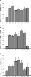
Relative mRNA expression of MpCTSK (A)/MpBCL-2 (B)/MpPO (C) in the hepatopancreases of M. petechialis at 0–13 dpi after immersion in V. parahaemolyticus by qRT-PCR. Error bars represent the SD. The asterisk (*) represents significant differences found when compared to 0 dpi (P < 0.05).
Figure 6
Figure 6
Antibacterial activities of rMpPO and rMpCTSK evaluated by the minimal inhibitory concentration method. (A,B) The inhibition of V. parahaemolyticus growth at 0, 8, 16, and 24 h post rMpPO- or rMpCTSK-addition. (C,D) The inhibition of Staphylococcus aureus growth at 0, 8, 16, and 24 h post rMpPO- or rMpCTSK-addition. Error bars represent the SD. Asterisks (*) represent a significant difference between groups given rMpPO/rMpCTSK and the control group (P < 0.05).
Figure 7
Figure 7
Antibacterial activities of rMpPO and rMpCTSK evaluated by the bacterial inhibition loop method. (A,B) The growth of V. parahaemolyticus on an LB agar plate with rMpPO or rMpCTSK added. (C,D) The growth of Staphylococcus aureus on an LB agar plate with rMpPO or rMpCTSK added. Kanamycin was used as positive control. GST-tagged protein/His-tagged protein and PBS were used as negative controls.
Figure 8
Figure 8
The apoptosis rate of haemocytes in the ABT199-treated group and the control group. Error bars represent the SD. Asterisks (*) represent a significant difference between the ABT199-treated group and the control group (P < 0.05).

References

    1. Hodgkinson CA, Moore KJ, Nakayama A, Steingrímsson E, Copeland NG, Jenkins NA, Arnheiter H. Mutations at the mouse microphthalmia locus are associated with defects in a gene encoding a novel basic-helix-loop-helix-zipper protein. Cell. (1993) 74:395–404. 10.1016/0092-8674(93)90429-T - DOI - PubMed
    1. Zhao GQ, Zhao Q, Zhou X, Mattei MG, de Crombrugghe B. TFEC, a basic helix-loop-helix protein, forms heterodimers with TFE3 and inhibits TFE3-dependent transcription activation. Mol Cell Biol. (1993) 13:4505–12. 10.1128/MCB.13.8.4505 - DOI - PMC - PubMed
    1. Beckmann H, Su LK, Kadesch T. TFE3: a helix-loop-helix protein that activates transcription through the immunoglobulin enhancer muE3 motif. Gene Dev. (1990) 4:167–79. 10.1101/gad.4.2.167 - DOI - PubMed
    1. Carr CS, Sharp PA. A helix-loop-helix protein related to the immunoglobulin E box-binding proteins. Mol Cell Biol. (1990) 10:4384–88. 10.1128/MCB.10.8.4384 - DOI - PMC - PubMed
    1. Hughes MJ, Lingrel JB, Krakowskyt JM, Anderson KP. A helix-loop-helix transcription factor-like gene is located at the mi locus. J Biol Chem. (1993) 268:20687–90. - PubMed

Publication types

MeSH terms

Substances

LinkOut - more resources