. 2019 May 23;7(5):e2271.
doi: 10.1097/GOX.0000000000002271. eCollection 2019 May.

Techniques, Principles and Benchmarks in Asian Blepharoplasty

Affiliations

Techniques, Principles and Benchmarks in Asian Blepharoplasty

William P D Chen. Plast Reconstr Surg Glob Open. .

Abstract

Background: Asian blepharoplasty is a deceptively simple procedure where the goal is to create an upper lid crease. The author presents a retrospective self-analysis of 362 cases performed over the past 12 years.

Methods: 362 cases that fits the inclusion criteria were tabulated into spreadsheet data format. Recorded were age, gender, date of service and follow-ups, whether the AB performed was for primary or revisional purpose; the preoperative lid crease status, the patient-chosen crease height as well as shape preferred. Intraoperative observation included presence or absence of preaponeurotic fat, whether partially resected, or reposited were noted.

Results: Of 362 patients (724 upper lids), primary AB constituted 81% (295) and revisional AB contributed 19% (67). The gender distribution was 87% female (315) and 13% male (47). The age distribution ranged from 12 to 75 years. The crease height selected ranged from 6.0 to 8.0 mm, with the median being 7.0 mm. Of the crease shape chosen, parallel shape was 65% (236) and nasally-joining crease shape was 35% (126).

Conclusions: Asian blepharoplasty via trapezoidal debulking of preaponeurotic platform is a safe, effective and anatomically-based technique that does not involve the use of permanent buried sutures. The article discussed the 5 essential factors (aponeurotic attachment, selective block clearance of preaponeurotic space, precise positioning of the crease formation loci, detection of latent droopy eyelids and avoidance of Faden-like suture effect) and the author's benchmarks to achieve a better success rate. Results for primary and revisional Asian blepharoplasty, strategies and potential pitfalls are presented. ( http://links.lww.com/PRSGO/B141).


Figures
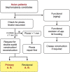
Fig. 1.
Fig. 1.
Clinical pathway showing selection of Asian blepharoplasty cases, both primary and revisional, in this series. (The yellow shaded zone highlights the group of included cases. All others were excluded.)
Video Graphic 1.
Video Graphic 1.
See video, Supplemental Digital Content 1, which displays Asian upper blepharoplasty. This video is available in the “Related Videos” section of the Full-Text article on PRSGlobalOpen.com or at http://links.lww.com/PRSGO/B79. Primary Double Eyelid Surgery—Trapezoidal Debulking of Upper Eyelid Tissues (Plastic and Reconstructive Surgery—Global Open. May 2018;6:e1780. doi:10.1097/GOX.0000000000001780).
Video Graphic 2.
Video Graphic 2.
See video, Supplemental Digital Content 2, which displays Asian upper blepharoplasty Part 2. This video is available in the “Related Videos” section of the Full-Text article on PRSGlobalOpen.com or at http://links.lww.com/PRSGO/B80. Revisional Asian Blepharoplasty—Bevelled Approach and Resetting of Eyelid Lamellas (Plastic and Reconstructive Surgery – Global Open. August 7, 2018;6:-e1785. doi:10.1097/GOX.0000000000001785).
Fig. 2.
Fig. 2.
A, Age distribution: the age distribution ranged from 12 to 75 years. The most common age group was from 21 to 30 years, although it spread across the 30- to 60-year-old spectrum. (Each bar includes a 5-year range that comprises those aged within 5 years up to that indicated age at the bottom of the bar.) B, Gender: 315 cases were women (87%), and 47 were for men (13%). C, There were 295 cases of primary Asian blepharoplasty (81%) and 67 cases for revisional Asian blepharoplasty (19%). D, Selection of crease shape: 65% (236) chose parallel crease shape, whereas 35% (126) chose nasally joining shape.
Fig. 3.
Fig. 3.
Upper lid crease height design. The crease height selected ranged from 6.0 to 8.0 mm, with the median being 7.0 mm (~49%). Slightly below-average crease height of 6.5 mm was the next most chosen height design, followed by slightly above-average crease height of 7.5 mm.
Fig. 4.
Fig. 4.
Treatment of preaponeurotic fat in primary Asian blepharoplasty (295 cases, 590 eyelids). Among these, 73% (green +yellow pies: 214 cases) had observable fat intraoperatively. Of these 214 cases with fat seen, 54% (115 cases; yellow) underwent partial reduction of preaponeurotic fat. Among 295 cases, 39% (115 cases) had underwent some form of fat reduction.
Fig. 5.
Fig. 5.
The crease construction is facilitated through placement of interrupted sutures, each directing a superficial portion of levator aponeurotic fibers along the superior tarsal border toward the upper and lower skin edges of the lid crease incision (Reprinted from “Chen WPD. Asian Blepharoplasty and the Eyelid Crease. 3rd ed. Elsevier Science; 2016”).
Fig. 6.
Fig. 6.
The concept of trapezoidal and triangular debulking of eyelid tissues as applied in Asian upper blepharoplasty. The beveled approach allows a selective removal of those tissues which may be impeding crease construction, and optimally aligns the wound for closure. After making the initial lid crease incision along the superior tarsal border (STB), plus an upper skin incision separated from it by 1.5–2 mm of skin, the upwardly-beveled trans-orbicularis arrow and returning vector (along the STB) represent the asymmetric sides of a conceptual trapezoid, with the skin and anterior surface of aponeurosis being the two remaining sides essentially running parallel to each other. Based on limited skin removal, the excision of soft tissues(skin, orbicularis, septum and fat) can be performed in an elegant, trapezoidal block. This permits a greater surface of the aponeurosis to be cleared (Reprinted from “Chen WPD. Asian Blepharoplasty and the Eyelid Crease. 3rd ed. Elsevier Science; 2016”).
Fig. 7.
Fig. 7.
Drawings illustrating the concept of Anatomic crease height represented by the purple line spanning the actual dimension of the tarsal plate; and Tilted crease height (Tch, in blue vertical) represents the vertical visual component of the tilted tarsus when observed frontally. When there is a lid fold partly shielding the crease, the segment of pretarsal skin we see exposed frontally is measured as the Apparent Crease height (green, vertical). Neither the Tch nor the apparent crease height represents the true extent of the anatomic crease height (Reprinted from “Chen WPD. Asian Blepharoplasty and the Eyelid Crease. 3rd ed. Elsevier Science; 2016”).
Fig. 8.
Fig. 8.
Treatment of incidental findings of medial upper eyelid fold. Undermining of redundant tissues followed by excision of orbicularis oculi, and overlapping dog-ears as applied in construction of a nasally joining crease shape (top 2 diagrams), and in parallel crease shape(lower two diagrams); note the deliberate placement of the medial-most suture knot inferior to the incision edge (down-knotting) for nasally joining crease shape, and on the upper border (up-knotting) for creating parallel crease shape (Reprinted from “Chen WPD. Asian Blepharoplasty and the Eyelid Crease. 3rd ed. Elsevier Science; 2016”)
Fig. 9.
Fig. 9.
Drawings showing placement and passages of permanent buried suture loops. a, Traditionally the suture methods uses three sets of double-armed 7-0 nylon or prolene. The drawing shows the typical passages for the central set of buried suture(left upper lid). The first passage (step 1) involves everting the upper lid margin and passing it subconjunctivally for 3–4 mm across (A′–B′) at a level typically several millimeters above the superior tarsal border, further up and behind the levator aponeurosis. The second passage (step 2) directs one needle toward the skin side along the path of B′–B, aiming just over the upper border of the tarsus. Similarly for the other arm of the suture, the third passage (step 3) goes from A′ to A. If each of the suture threads are tied on the skin at this moment, it will be a full thickness compression ligature encompassing Muller’s muscle, levator aponeurosis and orbicularis oculi muscle in a postero-superiorly biased fashion along the axis of levator muscle’s contractility. In addition to a plication effect on the aponeurosis, it inadvertently creates a “Faden-like effect” at each of the two locations of B′–B and A′–A. In actuality the second needle exiting the skin at A (after step 3) is re-passed (step 4) subcutaneously across to join B, exiting at a mini-stab skin opening there. The nylon ends are “firmly tied” and the knot sunken into the small surgical opening. In addition to the Faden-like effect, this results in a horizontal contraction in the width of levator aponeurosis at the two locations of A′–B′ and A–B. With three sets of sutures, the restrictive effect is tripled (Reprinted from “Chen WPD. Asian Blepharoplasty and the Eyelid Crease. 3rd ed. Elsevier Science; 2016”). Right, b, Cross section showing placement of buried suture that encircles the orbicularis oculi, levator aponeurosis and underlying Mueller’s muscle. Here it is shown as a blue 7-0 nonabsorbable suture loop. The nanospheres mentioned in the “Critical Crease-positioning” section is shown as a magnified yellow nanosphere here, is constrained in its phasic-reversal function; as the linkage of different layers added load to the levator when it contracts (causing ecto-Faden-like weakening effect for the levator on the long term), and tethering the anterior orbicularis to a higher point on the posterior levator muscle (one can say that here it is creating endo-Faden-like effect for the orbicularis oculi). The orbicularis oculi normally functions to shut the eyelid fissure, therefore locking it together with levator, which is an elevator/opening muscle makes the orbicularis work harder to close the lid fissure (Reprinted from “Chen WPD. Asian Blepharoplasty and the Eyelid Crease. 3rd ed. Elsevier Science; 2016”).
Video Graphic 3.
Video Graphic 3.
See video, Supplemental Digital Content 3, which demonstrates higher-than-average crease placement and its restrictive (impeding) effect on levator muscle excursion, in a routine functional upper blepharoplasty for a 49-y-old female. This video is available in the “Related Videos” section of the Full-Text article on PRSGlobalOpen.com or at http://links.lww.com/PRSGO/B81.
Video Graphic 4.
Video Graphic 4.
See video, Supplemental Digital Content 4, which displays surgical steps involved in medial rectus recession coupled with linking the muscle to adjacent orbital fat and connective tissues, in a 3-y-old child with congenital esotropia. It achieved similar impeding effect as posterior fixation with Faden suturing technique, but without the need for placement of intrascleral sutures and its associated risks. This video is available in the “Related Videos” section of the Full-Text article on PRSGlobalOpen.com or at http://links.lww.com/PRSGO/B82 (Edited from video, courtesy of Robert Clark M. D. of Long Beach, Calif.).
Fig. 10.
Fig. 10.
Pre- and postoperative views. A, 42-y-old female for primary Asian blepharoplasty, who preferred nasally joining crease shape, and medium crease height of 7 mm. B, 36-y-old female who presented with crease asymmetry of left eye 8.5 mm crease height, right eye 5.0 mm crease height (left column: preoperative); preferred staying with parallel shape, and chose 7.5 mm crease height (right column: postoperative views). C, 40-y-old female has permanent eyeliner, with crease height of 8.5 mm and pretarsal skin wrinkling; prefer stay with parallel shape and set to 7.5 mm crease height (right eye shown here). D, 29-y-old female for primary Asian blepharoplasty. Preferred medium crease height of 7.0 mm, and parallel crease shape (left eye shown here).

References

    1. Chen WP. Literature on double eyelid surgeries: appendix 1,2,3. In: Asian Blepharoplasty and the Eyelid Crease. 2016:3rd ed Edinburgh/New York: Elsevier; 345–365.
    1. Chen WP. Concept of triangular, trapezoidal, and rectangular debulking of eyelid tissues: application in Asian blepharoplasty. Plast Reconstr Surg. 1996;97:212–218. - PubMed
    1. Collin JR, Beard C, Wood I. Experimental and clinical data on the insertion of the levator palpebrae superioris muscle. Am J Ophthalmol. 1978;85:792–801. - PubMed
    1. Cheng J, Xu FZ. Anatomic microstructure of the upper eyelid in the oriental double eyelid. Plast Reconstr Surg. 2001;107:1665–1668. - PubMed
    1. Morikawa K, Yamamoto H, Uchinuma E, et al. Scanning electron microscopic study on double and single eyelids in Orientals. Aesthetic Plast Surg. 2001;25:20–24. - PubMed