

Icariin-mediated activation of autophagy confers protective effect on rotenone induced neurotoxicity in vivo and in vitro
- PMID: 31334034
- PMCID: PMC6624214
- DOI: 10.1016/j.toxrep.2019.06.014
Icariin-mediated activation of autophagy confers protective effect on rotenone induced neurotoxicity in vivo and in vitro
Erratum in
-
Erratum regarding missing Declaration of Competing Interest statements in previously published articles.Toxicol Rep. 2020 Dec 22;8:28-29. doi: 10.1016/j.toxrep.2020.12.007. eCollection 2021. Toxicol Rep. 2020. PMID: 33384945 Free PMC article.
Abstract
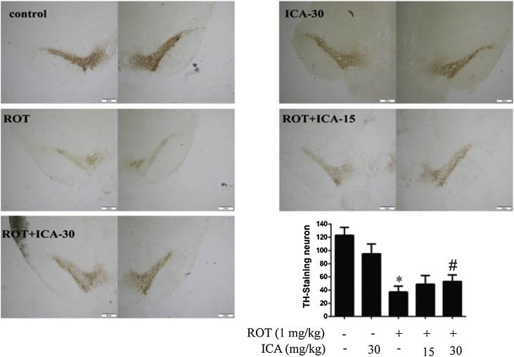
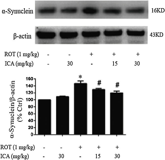
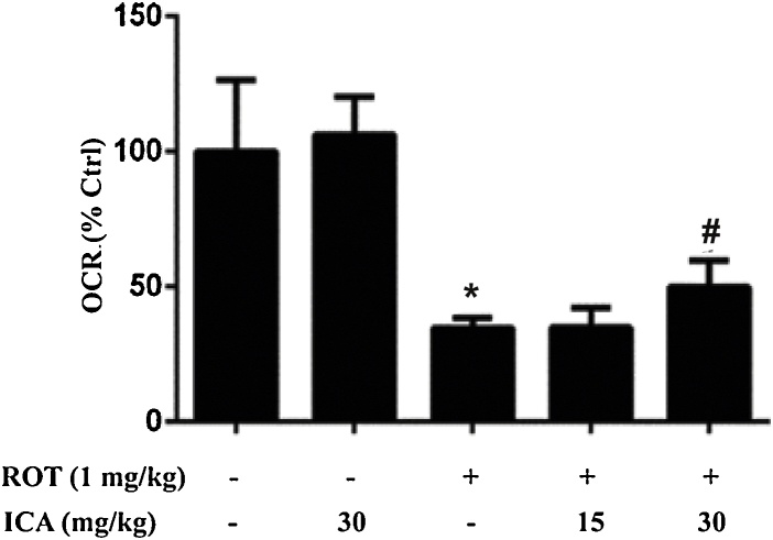
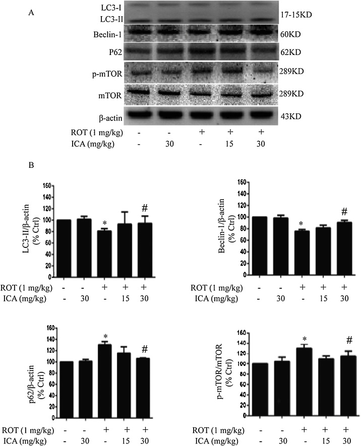
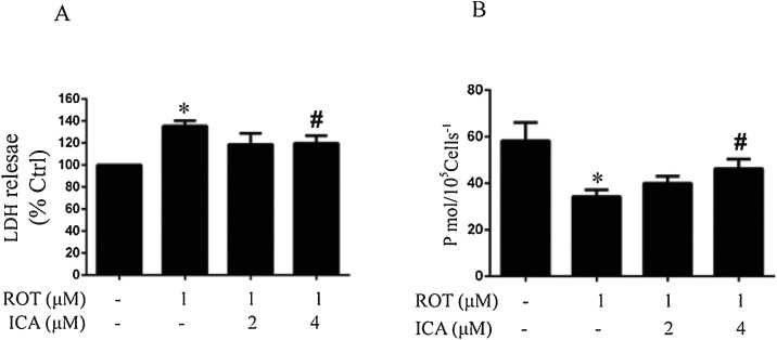
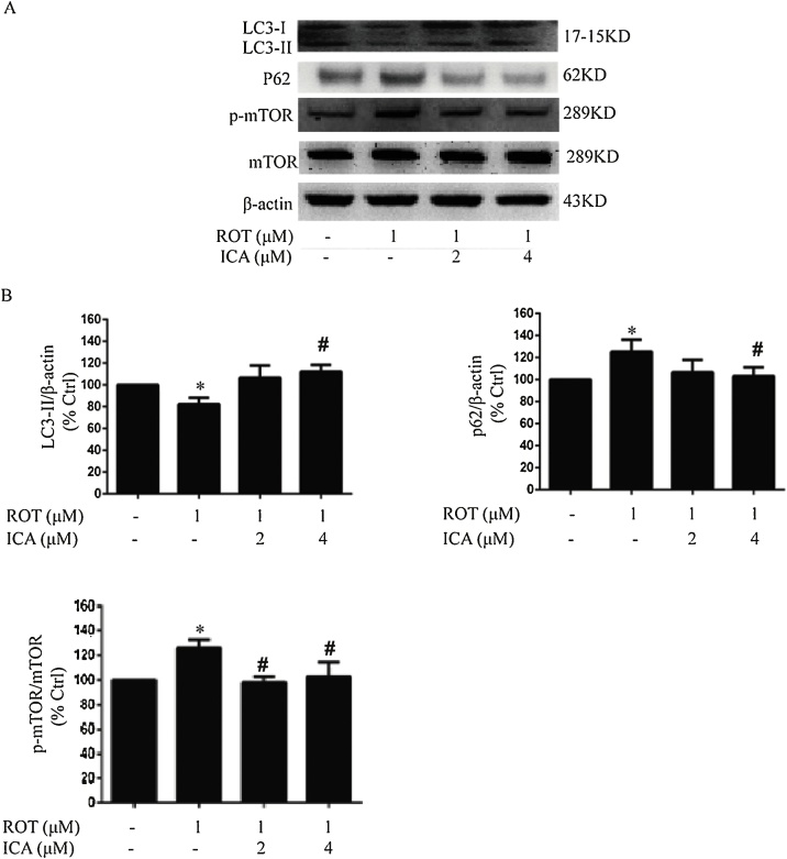
Rotenone (ROT) is an environmental neurotoxin which has been demonstrated to cause characteristic loss of dopamine (DA) neurons in Parkinson's disease (PD). Icariin (ICA) is a flavonoid glucoside isolated from Herba Epimedii that has been shown to display neuroprotective functions. The present study evaluated protective effects of ICA on ROT-induced neurotoxicity and determined the modulation of ICA on the regulation of autophagy in vivo and in vitro. Rats were treated with ROT (1.0 mg/kg/day) with a co-administration of ICA (15 or 30 mg/kg/day) for 5 weeks. Immunohistochemical analysis showed a significant loss in DA neurons in the substantia nigra (SN) of rats treated with ROT, accompanied by an increase in the accumulation of α-synuclein and a compromised mitochondrial respiration. However, co-administration of ICA potently ameliorated the ROT-induced neuronal cell injury and improved mitochondrial function and decreased the accumulation of α-synuclein. ROT treatment resulted in a decrease in the protein expression of LC3-II and Beclin-1, and an increase in the protein level of P62, and upregulated the activation of mammalian target of rapamycin (mTOR), whereas ICA significantly reversed these aberrant changes caused by ROT. Furthermore, the neuroprotective effect of ICA was further verified in PC12 cells. Cells treated with ROT displayed an increased cytotoxicity and a decreased oxygen consumption which were rescued by the presence of ICA. Furthermore, ROT decreased the protein expression level of LC3-II, enhanced Beclin-1 expression, and activated phosphorylation of mTOR, whereas ICA markedly reversed this dysregulation of autophagy caused by ROT in the PC12 cells. Collectively, these results suggest that ICA mediated activation of autophagic flux confers a neuroprotective action on ROT-induced neurotoxicity.
Keywords: Autophagy; BCA, bicinchoninic acid; DA, dopamine; DMEM, Dulbecco's modified Eagle's medium; HRP, horseradish peroxidase; ICA, icariin; Icariin; LDH, lactate dehydrogenase; Mitochondrial function; Neurotoxicity; OCR, oxygen consumption rate; PD, Parkinson`s disease; PE, phosphatidylethano-lamine; ROT, rotenone; Rotenone; SN, substantia nigra; mTOR, mammalian target of rapamycin.
Figures
References
-
- Dick F.D., Palma G.D., Ahmadi A., Scott N.W., Prescott G.J., Bennett J., Semple S., Dick S., Counsell C., Mozzoni P., Haites N., Wettinger S.B., Mutti A., Otelea M., Seaton A., Söderkvist P., Felice A., Geoparkinson Study Group Environmental risk factors for Parkinson’s disease and parkinsonism: the Geoparkinson study. Occup. Environ. Med. 2007;64(10):673–680. - PMC - PubMed
-
- O’Neal S.L., Zheng W. Manganese toxicity upon overexposure: a decade in review. Curr. Environ. Health Rep. 2015;2(3):315–328. https://doi.org/10.1007/s40572-015-0056-x. - PMC - PubMed
-
- Tanner C.M., Kamel F., Ross G.W., Hoppin J.A., Goldman S.M., Korell M., Marras C., Bhudhikanok G.S., Kasten M., Chade A.R., Comyns K., Richards M.B., Meng C., Priestley B., Fernandez H.H., Cambi F., Umbach D.M., Blair A., Sandler D.P., Langston J.W. Rotenone, paraquat, and Parkinson’s disease. Environ. Health Perspect. 2011;119(6):866–872. - PMC - PubMed
LinkOut - more resources
Full Text Sources
Miscellaneous
