A Rare Case of Possible Vacuolar Degeneration of Leprosy in Brain with Segmental Necrotizing Granulomatous Neuritis and Horner's Syndrome
- PMID: 31334067
- PMCID: PMC6615378
- DOI: 10.4103/idoj.IDOJ_388_18
A Rare Case of Possible Vacuolar Degeneration of Leprosy in Brain with Segmental Necrotizing Granulomatous Neuritis and Horner's Syndrome
Abstract
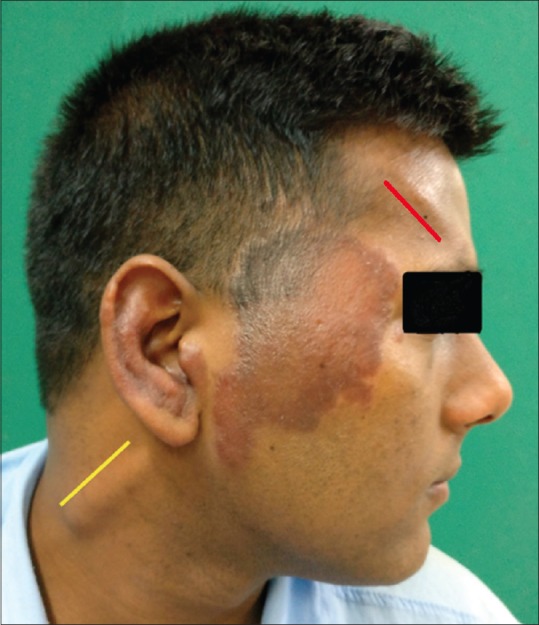
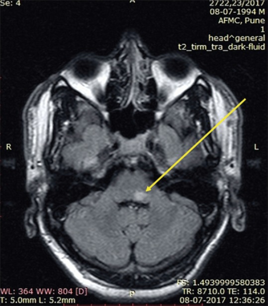
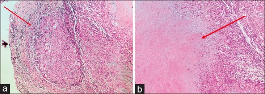
Leprosy has a predilection for peripheral nerves, but rarely does it involve the central nervous system (CNS). There is a single study of CNS involvement in leprosy showing vacuolar changes of motor neurons in medulla oblongata and spinal cord in autopsy findings. Besides this, there has been only one case report providing direct histopathological and molecular evidence of CNS involvement by leprosy in a living patient. Segmental necrotizing granulomatous neuritis (SNGN) is a rare condition affecting the peripheral nerves in leprosy usually seen as a complication of tuberculoid (TT) and borderline tuberculoid (BT) leprosy. We report the case of a 23-year-old male patient, a case of Hansen's disease (BT) who developed CNS involvement in the form of partial Horner's syndrome (right) and SNGN while on treatment. Magnetic resonance imaging of the brain revealed T2 hyperintense lesion on the dorsal aspect of left pontomedullary junction, suggestive of vacuolar degeneration of leprosy. Histopathology of greater auricular nerve (right) revealed SNGN.
Keywords: Granulomatous neuritis; Horner's syndrome; leprosy; segmental necrotizing; vacuolar degeneration.
Conflict of interest statement
There are no conflicts of interest.
Figures
References
-
- Aung T, Kitajima S, Nomoto M, En J, Yonezawa S, Arikawa I, et al. Mycobacterium leprae in neurons of the medulla oblongata and spinal cord in leprosy. J Neuropathol Exp Neurol. 2007;66:284–94. - PubMed
-
- Lee KH, Moon KS, Yun SJ, Won YH, Lee JH, Lee MC, et al. Brain involvement by leprosy presenting as a frontal cystic lesion. J Neurosurg. 2014;121:184–8. - PubMed
-
- Chandi SM, Chacko CJG, Fritschi EP, Job CK. Segmental necrotizing granulomatous neuritis of leprosy. Int J Lepr Other Mycobact Dis. 1980;48:41–7. - PubMed
-
- Vaidya MC, Palmer E, Weddell G, Rees RJ. A note on the presence of Mycobacterium leprae in the central nervous system of a mouse with lepromatous leprosy. J Med Microbiol. 1970;3:194–6. - PubMed