

Pearls and paradigms in Infective Keratitis
- PMID: 31334389
- PMCID: PMC6626937
Pearls and paradigms in Infective Keratitis
Abstract
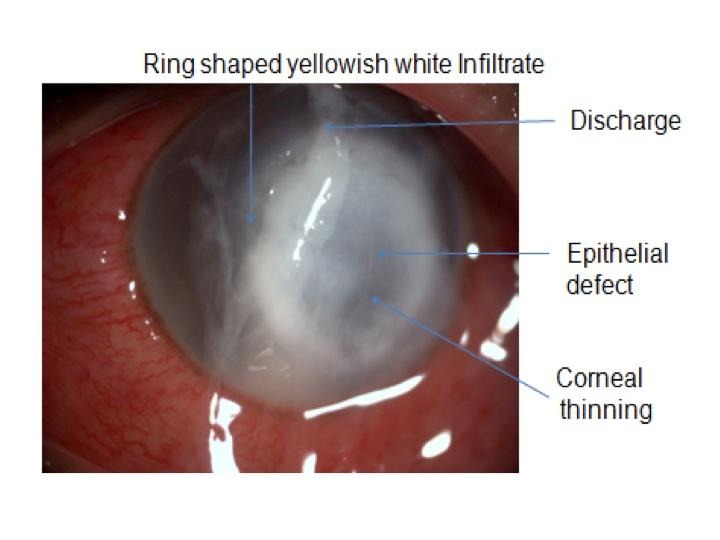
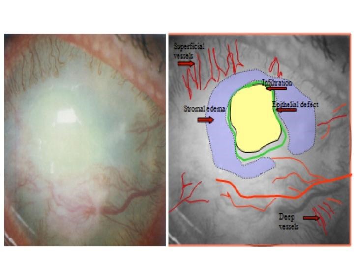
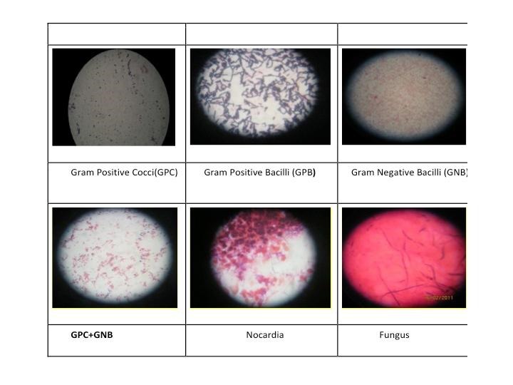
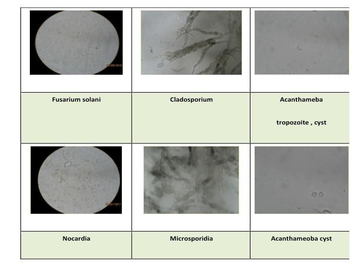
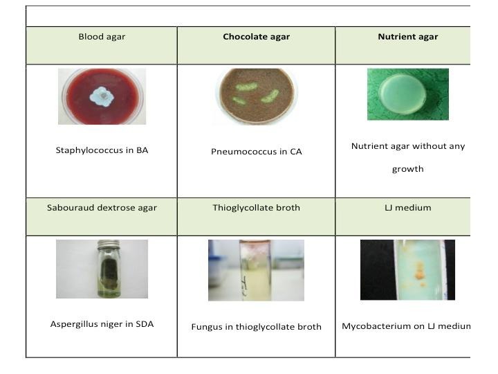
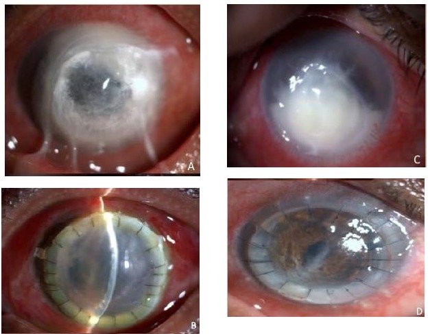
Infective Keratitis is a commonly encountered sight-threatening ocular emergency. In developing countries, it is a major cause of corneal blindness. Empirical treatment without microbiological work up often leads to treatment failure. Indiscriminate use of steroid antibiotic combination worsens the situation and makes further management challenging. The correct line of management can be potentially sight saving for both the ophthalmologist and the patient. This article on Infective keratitis has been written keeping best practices and protocols in mind. In a very simple and concise form, it focuses on the salient features of clinical presentation of infective keratitis and the stepwise approach to subsequent management in a patient. It explains in detail the way to perform corneal scraping, the importance of the same and further management based on microbiologically proven result. The management part includes indications and methods for medical as well as surgical intervention. We aimed to share our experience in the management of patients presenting with infective keratitis in the clinic.
Keywords: bacterial; culture; infective keratitis; management; microbiology; staining; treatment.
Figures
References
-
- McClellan KA. Mucosal defense of the outer eye. Surv Ophthalmol. 1997;42:233–246. - PubMed
-
- Dua HS, Gomes JA, Donoso LA, et al. The ocular surface as part of the mucosal immune system: conjunctival mucosa-specific lymphocytes in ocular surface pathology. Eye. 1995;9:261–267. - PubMed
-
- Ogawa GSH, Hyndiuk RA. In: Smolin G, Thoft RA (Eds). The Cornea Scientific Foundations and Clinical Practice. Bacterial Keratitis and Conjunctivitis, Chapter 5. 3rd ed. Philadelphia: Philadelphia; p. 125.
Publication types
MeSH terms
Substances
LinkOut - more resources
Full Text Sources
Medical