Review
. 2019 Sep:117:109168.
doi: 10.1016/j.biopha.2019.109168. Epub 2019 Jul 1.

The impact of infection and inflammation in oncologic 18F-FDG PET/CT imaging

Affiliations
Review

The impact of infection and inflammation in oncologic 18F-FDG PET/CT imaging

W Tania Rahman et al. Biomed Pharmacother. 2019 Sep.

Abstract

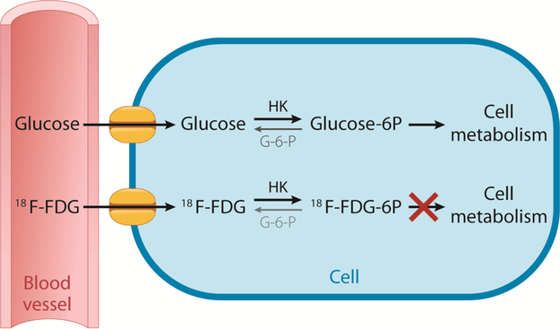
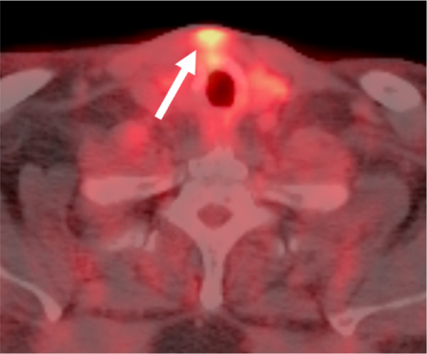
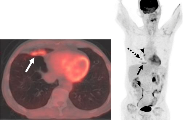
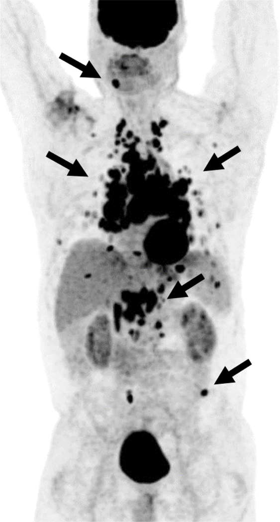
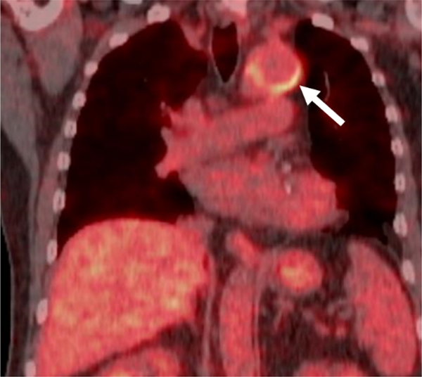
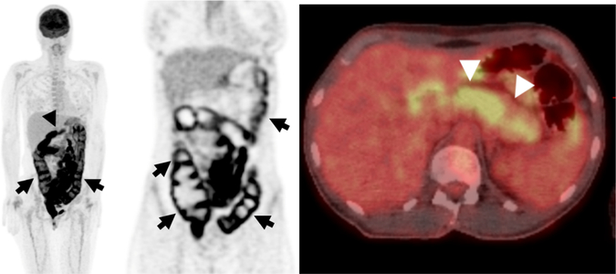
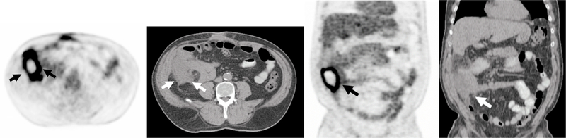
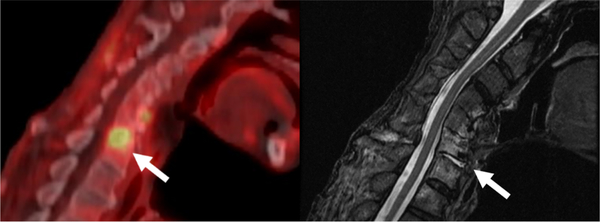
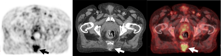
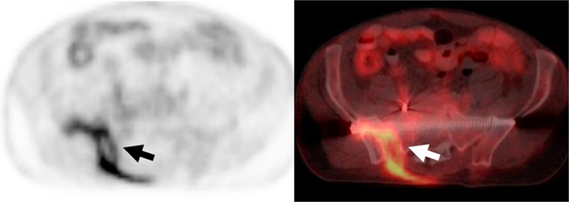
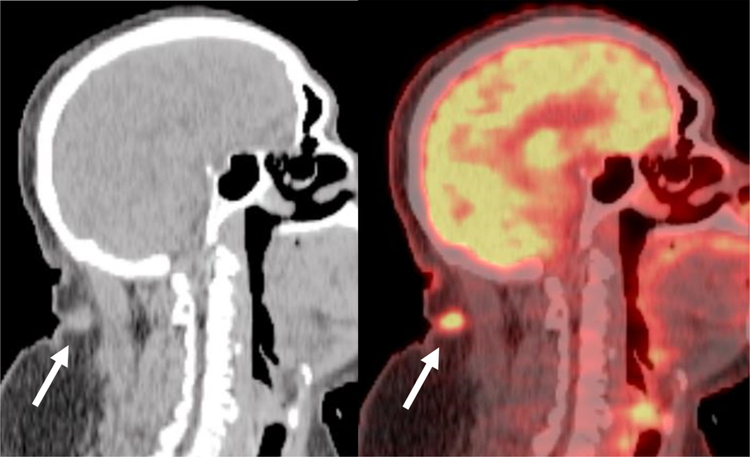
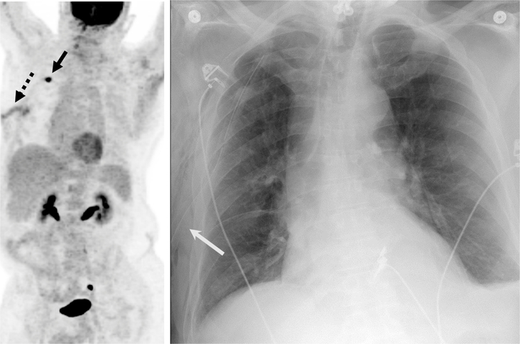
Sites of infection and inflammation can be misleading in oncology PET/CT imaging because these areas commonly show 18F-FDG activity. Caution in the interpretation must be taken to avoid the misdiagnosis of malignancy. Utilization of both CT findings as well as patient history can help differentiate benign infectious and inflammatory processes from malignancy, although occasionally additional work-up may be required. This article discusses the mechanism of 18F-FDG uptake in infection and inflammation with illustrative examples.

Keywords: (18)F-FDG PET/CT; Imaging pitfalls; Infection; Inflammation.


Figures

Fig. 1.
Fig. 1.
Glucose and 18F-FDG Cellular Uptake and Metabolism. Both glucose and 18F-FDG enter the cells by glucose transporter membrane proteins. Once in the cell, both are phosphorylated by hexokinase (HK), which can be reversed by glucose-6-phosphatase (G-6-P) if present. Phosphorylated glucose (Glucose-6 P) is available to be further metabolized by the cell, whereas phosphorylated 18F-FDG (18F-FDG-6 P) cannot be further metabolized and is therefore considered “trapped”.
Fig. 2.
Fig. 2.
Focal Inflammatory 18F-FDG uptake at Site of a Previous Tracheostomy. Axial fused PET/CT demonstrates focal uptake in the soft tissues (arrow) in the anterior neck superficial to the thyroid gland that correlated to the site of postsurgical inflammation at the site of a previous tracheostomy.
Fig. 3.
Fig. 3.
Postobstructive 18F-FDG-avid Airspace Disease in a Patient with Lung Malignancy. A, B Axial fused PET/CT and maximum intensity projection image demonstrates focal uptake in right middle lobe airspace disease (solid arrows) in a patient with a hypermetabolic central right lung nodule (dashed arrow) and an adjacent right hilar lymph node (arrowhead).
Fig. 4.
Fig. 4.
Multiple Foci of Abnormal Uptake in a 56-year-old male with 18F-FDG-avid Sarcoidosis. Maximum intensity projection image from an 18F-FDG PET/CT multiple hypermetabolic foci (arrows) in lymph nodes, pulmonary nodules, as well as several osseous foci that were initially viewed with concern for a malignancy such as lymphoma. However, subsequent tissue sampling revealed sarcoidosis as the underlying cause.
Fig. 5.
Fig. 5.
Incidental Non-infectious Inflammatory Uptake in a Recently Placed Thoracic Aortic Graft in a 59-year-old male. Fused coronal 18F-FDG PET/CT demonstrates mild intensity curvilinear uptake (arrow) along portions of a recently placed thoracic aortic graft in an oncology patient without signs/symptoms of infection.
Fig. 6.
Fig. 6.
42-year-old Woman Receiving Immunotherapy for Metastatic Melanoma. A, Maximum intensity projection image from an 18F-FDG PET/CT demonstrates diffuse hypermetabolic activity in the colon (arrows) and diffuse uptake involving the pancreas (arrowhead). B, Coronal PET images show diffuse uptake throughout the entire colon consistent with colitis (arrows), an adverse effect related to immunotherapy.

C, Axial fused PET/CT show diffuse uptake throughout the pancreas (arrowheads) consistent with pancreatitis related to immunotherapy.
Fig. 7.
Fig. 7.
Hypermetabolic Right-sided Abdominal Abscess 64 year-old Man. A, B Axial and C, D coronal PET and CT images from an 18F-FDG PET/CT demonstrates a hypermetabolic fluid collection with peripheral uptake and central photopenia in the right abdomen (arrows). The patient had recent colonoscopy and biopsy of a polyp at the cecum with suspicion for perforation after the procedure. The abscess required to surgical drainage and antibiotic treatment.
Fig. 8.
Fig. 8.
67-year-old Male with Focal 18F-FDG uptake correlating to Cervical Discitis/Osteomyelitis. A, Fused sagittal 18F-FDG PET/CT demonstrates focal uptake at C6-C7 (arrow) concerning for infection. B, Sagittal T2 weighted images demonstrates focal edema/fluid in the C6-C7 disc space and adjacent vertebral bodies (arrows) concerning for discitis/osteomyelitis. Patient’s spinal infection was determined to be a complication of his treatment for his head and neck cancer.
Fig. 9.
Fig. 9.
72-year-old Man with Sacral Decubitus Ulcer. A-C, Axial PET, CT and fused from an 18F-FDG PET/CT demonstrates focal uptake within an ulcer with extension to the involve the sacrum consistent with infected sacral decubitus ulcer and sacral osteomyelitis.
Fig. 10.
Fig. 10.
Hypermetabolic Septic Arthritis Incidentally Detected in on 18F-FDG PET/CT performed for a Solitary Pulmonary Nodule Evaluation. A, B, Axial 18F-FDG PET and fused PET/CT demonstrates curvilinear uptake in the right sacroiliac joint and surrounding soft tissues (arrows) corresponding to a septic joint with a sinus tract. The patient had a history of multiple pelvic surgeries due to prior trauma.
Fig. 11.
Fig. 11.
72-year-old male with Lung Cancer with Incidental Focal Uptake in the Neck. A, B, Sagittal CT and fused 18F-FDG PET/CT of the neck demonstrates a small subcutaneous hypermetabolic nodule in the posterior soft tissues of the neck (arrow). Clinically this site corresponded to a furuncle (boil) with adjacent skin erythema.
Fig. 12.
Fig. 12.
67-year-old man with a Recent Diagnosis of Lung Carcinoma. A, Maximum intensity projection image from an 18F-FDG PET/CT demonstrates a hypermetabolic right upper lobe mass (solid black arrow) with an additional linear focus of uptake in the subcutaneous tissues of the right chest wall (dashed arrow) that was inflammatory due to a right thoracotomy tube placed due to a post-biopsy pneumothorax. B, Frontal chest radiograph demonstrates the location of the right thoracotomy tube (solid white arrow) confirming the inflammatory etiology of the FDG uptake.

References

    1. Safaie E, Matthews R, Bergamaschi R, PET scan findings can be false positive, Tech. Coloproctol 19 (6) (2015) 329–330. - PubMed
    1. Annibaldi A, Widmann C, Glucose metabolism in cancer cells, Curr. Opin. Clin. Nutr. Metab. Care 13 (4) (2010) 466–470. - PubMed
    1. Gallagher BM, Fowler JS, Gutterson NI, MacGregor RR, Wan CN, Wolf AP, Metabolic trapping as a principle of oradiopharmaceutical design: some factors resposible for the biodistribution of [18F] 2-deoxy-2-fluoro-D-glucose, J. Nucl. Med 19 (10) (1978) 1154–1161. - PubMed
    1. Smith TA, FDG uptake, tumour characteristics and response to therapy: a review, Nucl. Med. Commun 19 (2) (1998) 97–105. - PubMed
    1. Vaidyanathan S, Patel CN, Scarsbrook AF, Chowdhury FU, FDG PET/CT in infection and inflammation–current and emerging clinical applications, Clin. Radiol 70 (7) (2015) 787–800. - PubMed

MeSH terms

Substances