. 2019 Sep;40(8):e774-e781.
doi: 10.1097/MAO.0000000000002313.

Density of Macrophages Immunostained With Anti-iba1 Antibody in the Vestibular Endorgans After Cochlear Implantation in the Human

Affiliations

Density of Macrophages Immunostained With Anti-iba1 Antibody in the Vestibular Endorgans After Cochlear Implantation in the Human

Tadao Okayasu et al. Otol Neurotol. 2019 Sep.

Abstract

Hypothesis: Cochlear implantation may result in an increase in the density of macrophages in vestibular endorgans in the human.

Background: Vestibular symptoms are a common complication of cochlear implantation. In a previous study, we demonstrated histological evidence of a foreign-body response caused by silicon and platinum in the human cochlea following cochlear implantation. The objective of the current study was to seek evidence of a possible immune response in vestibular endorgans after cochlear implantation.

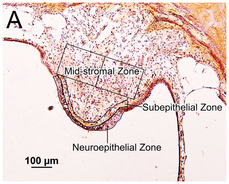
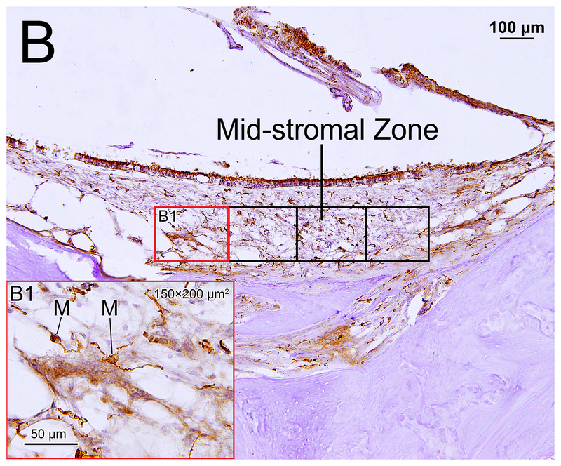
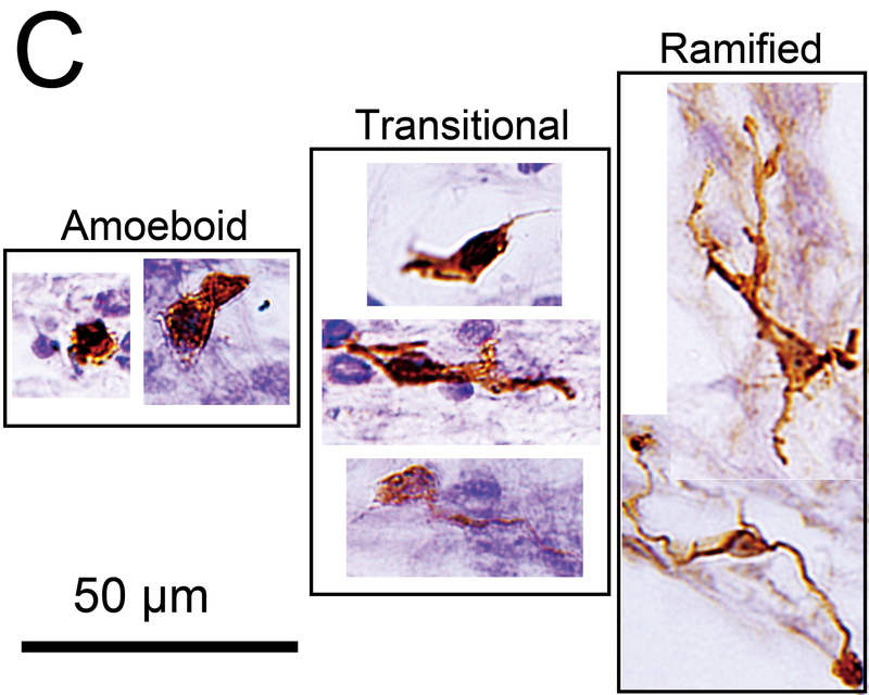
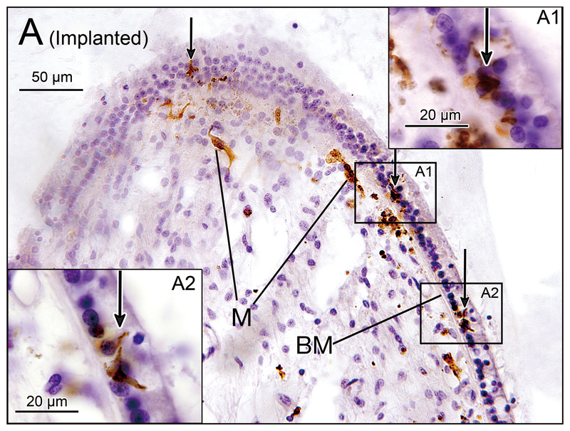
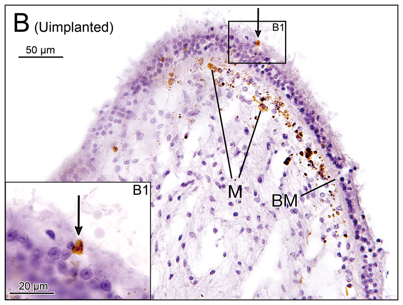
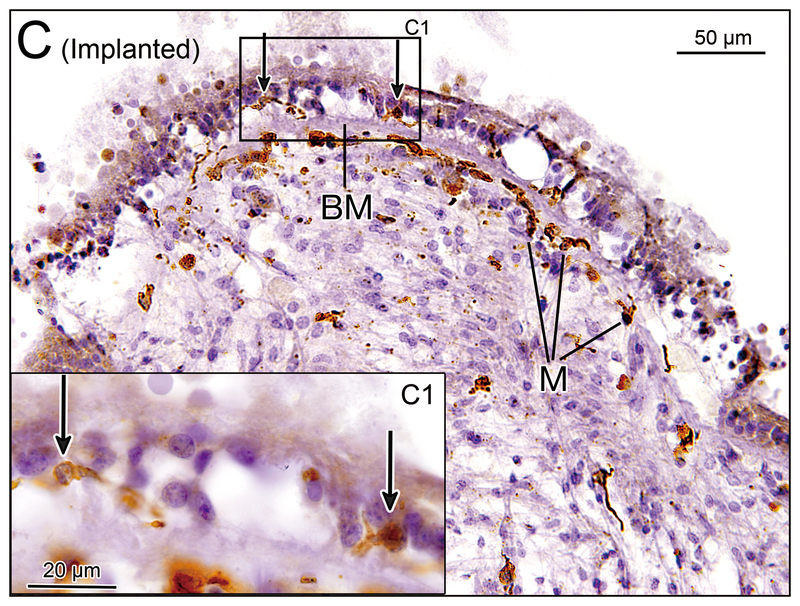
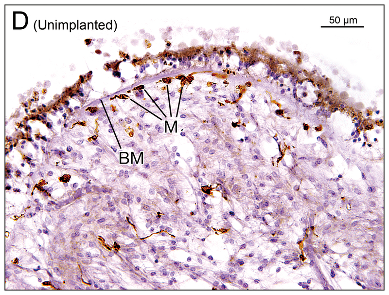
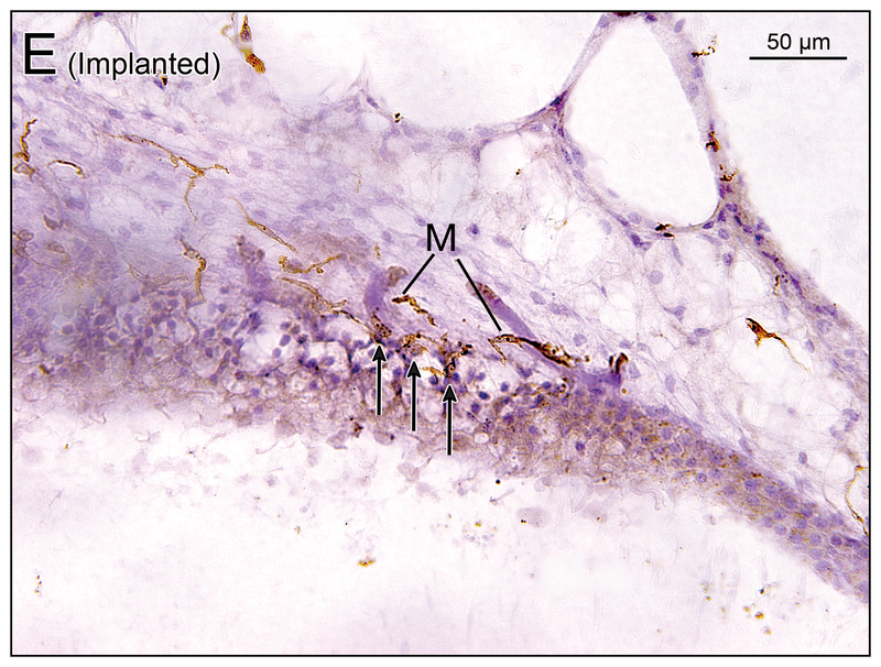
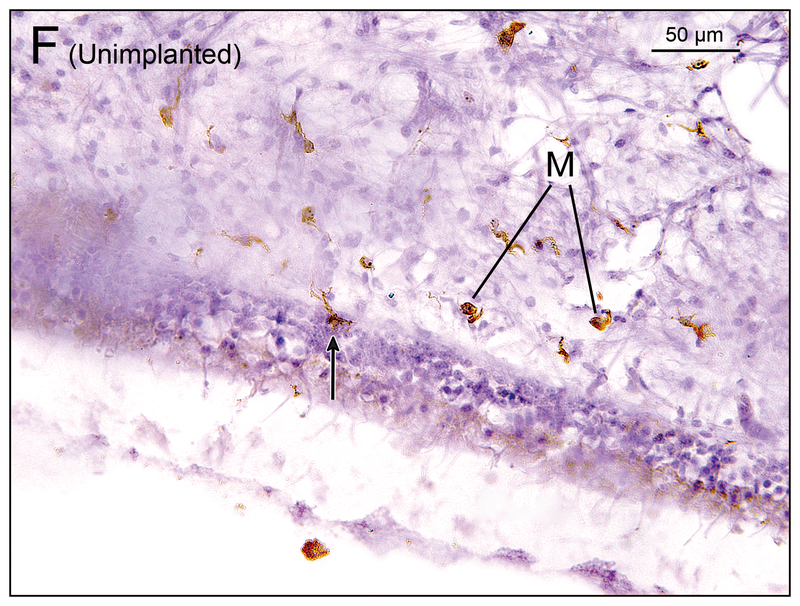
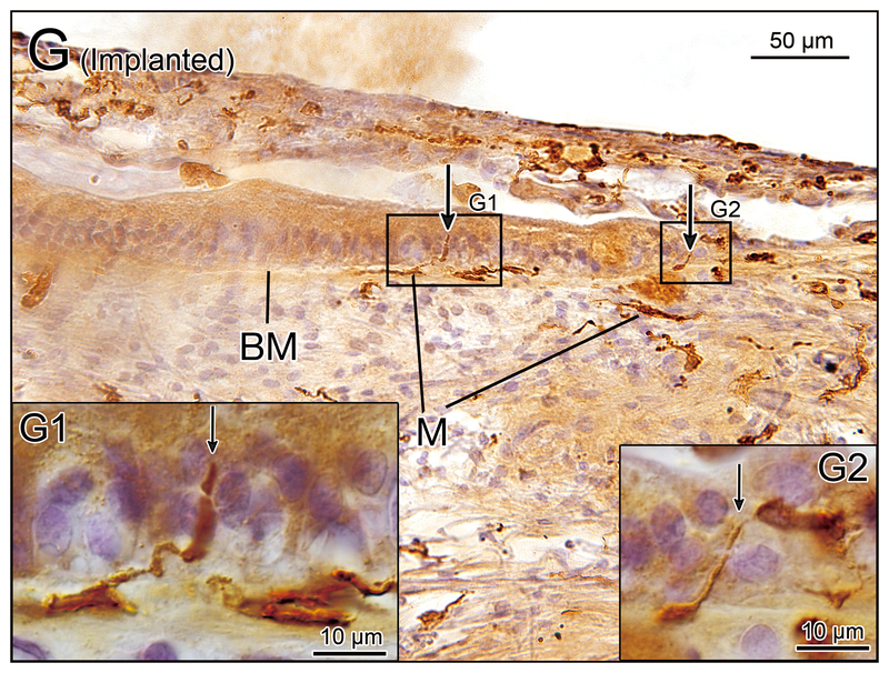
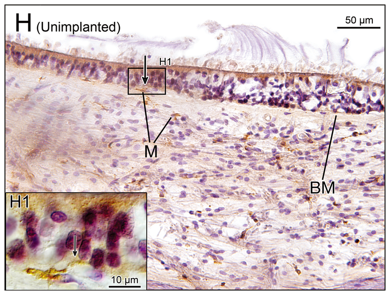
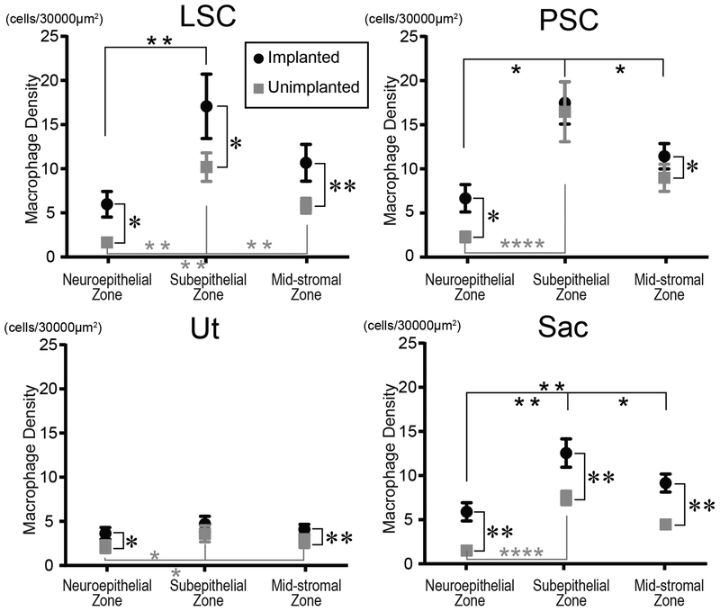
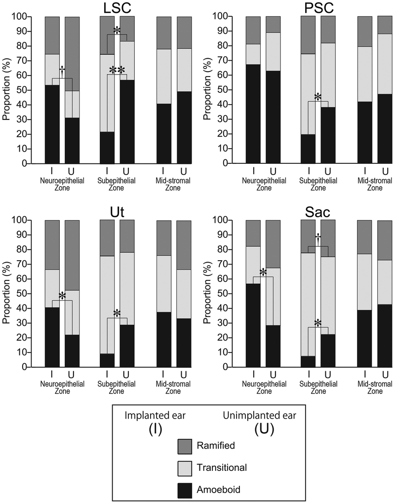
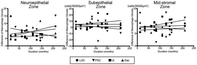
Methods: The density of macrophages immunostained with anti-Iba1 antibody in the vestibular endorgans (lateral and posterior semicircular canals, utricle and saccule) in 10 human subjects who had undergone unilateral cochlear implantation was studied by light microscopy. The densities of macrophages in the neuroepithelium, subepithelial stroma, and among dendritic processes in the mid-stromal zone in four vestibular endorgans in the implanted and the opposite unimplanted ears were compared. The distributions of macrophage morphology (amoeboid, transitional and ramified) were also compared.

Results: The densities of macrophages in implanted ears in four vestibular endorgans were significantly greater than that in opposite unimplanted ears except in the subepithelial zone of the utricle and posterior semicircular canal. In contrast to the neuroepithelium, the subepithelial distribution of amoeboid macrophages in implanted ears was significantly less than in unimplanted ears.

Conclusion: An increase in the density of macrophages in four vestibular endorgans after implantation was demonstrated. The transition among phenotype of macrophages suggested possible migration of amoeboid macrophages from the subepithelial stroma into the neuroepithelium.


Conflict of interest statement

(disclosure)

All authors declare no conflict of interest related to this manuscript.

Figures

Figure 1.
Figure 1.
Iba1- immunostaining of vestibular endorgans: quantification and subtyping of macrophages. (A) Crista ampularis of posterior semicircular canal of unimplanted right ear in case 7. The neuroepithelial and subepithelial zones are outlined. The areas studied in the mid-stromal zone are outlined into the two boxes, each boxed area measuring 150×200 μm2. The densities of Iba1-positive macrophages in these areas were calculated. (B) Saccule of implanted left ear in case 2. The stromal areas studied within the mid-stromal zone are outlined by the six boxes. (B1) A higher magnification of left red box seen in (B) is shown. Immunostained macrophages (M) were seen. (C) Macrophage subtypes. The amoeboid type has few processes and a relatively round cell body with a large nucleus. The ramified type has long branching processes. The transitonal type displays intermediate morphology.
Figure 1.
Figure 1.
Iba1- immunostaining of vestibular endorgans: quantification and subtyping of macrophages. (A) Crista ampularis of posterior semicircular canal of unimplanted right ear in case 7. The neuroepithelial and subepithelial zones are outlined. The areas studied in the mid-stromal zone are outlined into the two boxes, each boxed area measuring 150×200 μm2. The densities of Iba1-positive macrophages in these areas were calculated. (B) Saccule of implanted left ear in case 2. The stromal areas studied within the mid-stromal zone are outlined by the six boxes. (B1) A higher magnification of left red box seen in (B) is shown. Immunostained macrophages (M) were seen. (C) Macrophage subtypes. The amoeboid type has few processes and a relatively round cell body with a large nucleus. The ramified type has long branching processes. The transitonal type displays intermediate morphology.
Figure 2.
Figure 2.
(A) Crista ampularis of the lateral semicircular canal of implanted left ear in case 4. Iba1-immunostaining of vestibular endorgans: implanted (A, C, E, G) and unimplanted (B, D, F, H) ears. There were several macrophages (M) immediately beneath the basement membrane (BM) and a few macrophages were seen in the neuroepithelium (arrows). (A1) and (A2) A higher magnification of boxes in (A). (B) Crista ampularis of the lateral semicircular canal of unimplanted right ear in case 4. There were several macrophages (M) seen beneath the basement membrane and one macrophage was seen in the neuroepithelium (arrow). (B1) A higher magnification of box in (B). (C) Crista ampularis of the posterior semicircular canal of implanted left ear in case 2. There were several macrophages (M) seen beneath the basement membrane (BM) and a few macrophages were seen in the neuroepithelium (arrows). [See (C1)]. (C1) A higher magnification of box in (C). (D) Crista ampularis of the posterior semicircular canal of unimplanted right ear in case 2. There were several macrophages (M) seen beneath the basement membrane (BM), and a few macrophages were seen in the neuroepithelium. (E) Utricle of implanted right ear in case 1. Many macrophages (M) were seen in the neuroepithelium (arrows). (F) Utricle of unimplanted left ear in case 1. There were some macrophages (M) in the subepithelial stroma and a single macrophage was seen in the neuroepithelium (arrow). (G) Saccule of implanted left ear in case 7. There were some macrophages (M) below the basement membrane (BM) in the subepithelial stroma and long processes of some macrophages extended into the neuroepithelium (arrows). [See (G1) and (G2)]. (G1) and (G2) A higher magnification of boxes in (G) by Nomarski (differential interference contrast) microscopy. (H) Saccule of unimplanted right ear in case 7. There were some macrophages (M) in the subepithelial stroma. There is a single macrophage below the basement membrane (BM) and long process of the macrophage extended into the neuroepithelium (arrow). [See (H1)] (H1) A higher magnification of box in (H) by Nomarski microscopy.
Figure 2.
Figure 2.
(A) Crista ampularis of the lateral semicircular canal of implanted left ear in case 4. Iba1-immunostaining of vestibular endorgans: implanted (A, C, E, G) and unimplanted (B, D, F, H) ears. There were several macrophages (M) immediately beneath the basement membrane (BM) and a few macrophages were seen in the neuroepithelium (arrows). (A1) and (A2) A higher magnification of boxes in (A). (B) Crista ampularis of the lateral semicircular canal of unimplanted right ear in case 4. There were several macrophages (M) seen beneath the basement membrane and one macrophage was seen in the neuroepithelium (arrow). (B1) A higher magnification of box in (B). (C) Crista ampularis of the posterior semicircular canal of implanted left ear in case 2. There were several macrophages (M) seen beneath the basement membrane (BM) and a few macrophages were seen in the neuroepithelium (arrows). [See (C1)]. (C1) A higher magnification of box in (C). (D) Crista ampularis of the posterior semicircular canal of unimplanted right ear in case 2. There were several macrophages (M) seen beneath the basement membrane (BM), and a few macrophages were seen in the neuroepithelium. (E) Utricle of implanted right ear in case 1. Many macrophages (M) were seen in the neuroepithelium (arrows). (F) Utricle of unimplanted left ear in case 1. There were some macrophages (M) in the subepithelial stroma and a single macrophage was seen in the neuroepithelium (arrow). (G) Saccule of implanted left ear in case 7. There were some macrophages (M) below the basement membrane (BM) in the subepithelial stroma and long processes of some macrophages extended into the neuroepithelium (arrows). [See (G1) and (G2)]. (G1) and (G2) A higher magnification of boxes in (G) by Nomarski (differential interference contrast) microscopy. (H) Saccule of unimplanted right ear in case 7. There were some macrophages (M) in the subepithelial stroma. There is a single macrophage below the basement membrane (BM) and long process of the macrophage extended into the neuroepithelium (arrow). [See (H1)] (H1) A higher magnification of box in (H) by Nomarski microscopy.
Figure 3.
Figure 3.
Density of macrophages in vestibular endorgans in the three zones (neuroepithelium, subepithelal stroma, and mid-stroma). Black circles and gray squares indicate implanted and unimplanted ears, respectively. LSC= lateral semicircular canal; PSC= posterior semicircular canal; Ut = utricle; Sac = saccule. Asterisks (*) indicate the statistical significance between implanted and unimplanted ear (*, p<0.05; **, p<0.01; paired-t test or Wilcoxon matched-pairs signed rank test) and asterisks (*) indicate the statistical significance among the three zones (*, p<0.05; **, p<0.01; ****, p<0.0001; one-way repeated measures ANOVA followed by Tukey’s multiple comparisons test or Friedman test followed by Dunn’s multiple comparisons test).
Figure 4.
Figure 4.
Distribution of macrophage morphology. The proportion of the three macrophage morphologies in the three zones of vestibular endorgans. Amoeboid, transitional and ramified types are shown. The columns of implanted ear (I) and unimplanted ear (U) are indicated side by side. The asterisks (*) indicate the statistical significance between implanted and unimplanted ears (*, p<0.05; **, p<0.01; paired-t test or Wilcoxon matched-pairs signed rank test). The daggers (†) indicate a significant tendency (†, 0.05≤ p<0.1).
Figure 5.
Figure 5.
Correlation between density of macrophages vs duration after implantation. Scatter plot of difference of macrophage density (implanted minus unimplanted) verses duration (months after cochlear implantation) in the three zones in the four vestibular endorgans. No significant correlations were found (Pearson’s or Spearman’s coefficients of correlation test).

Similar articles

Cited by

References

    1. Fina M, Skinner M, Goebel JA, Piccirillo JF, Neely JG. Vestibular dysfunction after cochlear implantation. Otol Neurotol. 2003, 24: 234–242. - PubMed
    1. Terry B, Kelt RE, Jeyakumar A. Delayed complications after cochlear implantation. JAMA Otolaryngol Head Neck Surg. 2015, 141: 1012–1017. - PubMed
    1. Ibrahim I, Da Silva SD, Segal B, Zeitouni A. Effect of cochlear implant surgery on vestibular function: Meta-analysis study. J Otolaryngol Head Neck Surg. 2017, 46: 44. - PMC - PubMed
    1. Ito J Influence of the multichannel cochlear implant on vestibular function. Otolaryngol Head Neck Surg. 1998, 118: 900–902. - PubMed
    1. Kubo T, Yamamoto KI, Iwaki T, Doi K, Tamura M. Different forms of dizziness occurring after cochlear implant. Eur Arch Otorhinolaryngol. 2001, 258: 9–12. - PubMed

Publication types