. 2020 Jun 11;12(5):337-344.
doi: 10.1093/jmcb/mjz087.

ZC4H2 stabilizes RNF220 to pattern ventral spinal cord through modulating Shh/Gli signaling

Affiliations

ZC4H2 stabilizes RNF220 to pattern ventral spinal cord through modulating Shh/Gli signaling

Pengcheng Ma et al. J Mol Cell Biol. .

Abstract

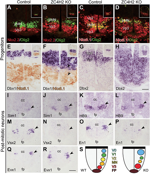
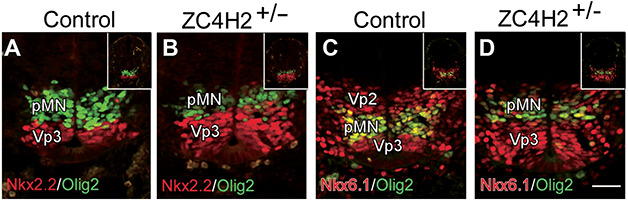
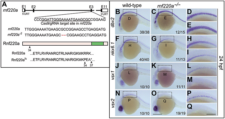
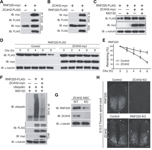
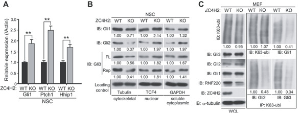
ZC4H2 encodes a C4H2 type zinc-finger nuclear factor, the mutation of which has been associated with disorders with various clinical phenotypes in human, including developmental delay, intellectual disability and dystonia. ZC4H2 has been suggested to regulate spinal cord patterning in zebrafish as a co-factor for RNF220, an ubiquitin E3 ligase involved in Gli signaling. Here we showed that ZC4H2 and RNF220 knockout animals phenocopy each other in spinal patterning in both mouse and zebrafish, with mispatterned progenitor and neuronal domains in the ventral spinal cord. We showed evidence that ZC4H2 is required for the stability of RNF220 and also proper Gli ubiquitination and signaling in vivo. Our data provides new insights into the possible etiology of the neurodevelopmental impairments observed in ZC4H2-associated syndromes.

Keywords: Gli signaling; RNF220; ZC4H2; patterning; spinal cord.


Figures

Figure 1
Figure 1
Developmental expression of ZC4H2 and construction of ZC4H2 knockout mice. (AE) Whole-mount in situ hybridization showing that ZC4H2 is expressed in the developing neural system at E8.5 (A), E9.5 (B and C), and E10.5 (D and E). (C) Section of E9.5 embryos at spinal cord shows that ZC4H2 is expressed in the ventral zone of spinal cord. (E) In E10.5 embryos, ZC4H2 expression is also detected in the limb buds. Scale bar, 400 μm in A, 300 μm in B, 50 μm in C, 700 μm in D, and 150 μm in E. (F) Diagram of the targeting construct and expected recombination events. Exon 2 of ZC4H2 is floxed by two Loxp sites. (G and H) Photographs showing control (G, WT) and ZC4H2−/− (H, KO) pups on the day of birth. The ZC4H2−/− pups die after birth. Scale bar, 5 mm (H, applies to G).
Figure 2
Figure 2
Progenitor and post-mitotic neuron domains are altered in the ventral spinal cord of the ZC4H2 knockout (KO) mice at E10.5. (A and B) The Nkx2.2+ (red) VP3 domain is expanded dorsally, while the Olig2+ (green) pMN domain is reduced in the ZC4H2 KO mice relative to controls. (C and D) Double labelling of Dbx1 and Nkx6.1 (red) and Olig2 (green) shows a dorsal shift and reduction of the Olig2+ pMN domain and the Vp2 domain was decreased in ZC4H2 KO spinal tube. (E and F) In situ immunostaining of Dbx1 and Nkx6.1 shows that the Dbx1+ Vp0 domain is increased and expanded ventrally to the dorsal boundary of the Nkx6.1+ domain in the ZC4H2 KO mice, whereas there is a gap region (i.e. Vp1 domain) between the Nkx6.1+ and Dbx1+ Vp0 domains in controls. (G and H) Dbx2 expression is expanded ventrally to the floor plate in ZC4H2 KO mice. Inserts in AH are low magnification pictures of the whole spinal tube. Scale bar, 50 μm (H, applies to AG). (I and J) The number of Sim1+ V3 neurons is dramatically increased in the ZC4H2 KO mice (J) compared with controls (I). Triangles point to their expressions in the ventral tube. (K and L) The number of Hb9+ MN neurons is reduced in the ZC4H2 KO mice (L) compared with controls (K). (M and N) Expression of Vsx2 is not detected in the ZC4H2 KO mice (N) but observed in control spinal cord (M). (O and P) The number of En1+ V1 neurons is greatly reduced in the ZC4H2 KO mice (P) compared with controls (O). (Q and R) The number of Evx1+ V0 neurons is increased in the ZC4H2 KO mice (R) compared with controls (Q). Triangles in I–R point to their expressions in the ventral tube. cc, central canal; fp, floor plate. Scale bar, 100 μm (R, applies to IQ). (S) A cartoon showing the relative changes of the different neuronal territories in the ZC4H2 KO ventral spinal cord. Note that there is no difference in the changes of the progenitor and neuronal domains between female ZC4H2−/− and male ZC4H2−/Y embryos (data not shown). The data shown here were all from ZC4H2−/− female embryos.
Figure 3
Figure 3
Changes of the progenitor domains in the ZC4H2 heterozygote (ZC4H2+/−) spinal cord at E10.5. (A and B) Double immunostaining shows that the Nkx2.2+ (red) Vp3 domain is expanded, while the Olig2+ (green) pMN domain is reduced along the D–V axis of the ventricular zone in ZC4H2+/− mice relative to controls. (C and D) Double immunostaining for Nkx6.1 (red) and Olig2 (green) shows a dorsal shift of the Nkx6.1+/Olig2+ pMN domain and a reduction of the Vp2 domain in the ZC4H2+/− spinal cord. Inserts in AD are low magnification pictures of the whole spinal tube. Scale bar, 50 μm (D, applies to AC).
Figure 4
Figure 4
Neural progenitors and post-mitotic neurons are altered in rnf220a knockout zebrafish embryos. (A) Schematic representation of CRISPR/Cas9-mediated genome modification. The black box represents the exon and the white box represents the non-coding region. The CRISPR/Cas9 target site is located in the third exon of rnf220a genomic locus, marked with a black underline. Lastly, a 2-bp deletion (labelled in red) induced by CRISPR/Cas9 results in a frame shift. Both wild-type Rnf220a and Rnf220fs proteins are shown. The wild-type protein is truncated to 37 amino acids by this mutation. fs, frame shit. (BI) Genes marking the progenitor domains of ventral spinal cord were examined by whole-mount in situ hybridization in rnf220a homozygous mutant (rnf220a−/−) embryos at 24 hpf. dbx2 gene expression domain is expanded ventrally (BE) at the expense of the reduced nkx6.1 expression domain (FI). (JQ) Whole-mount in situ hybridization analysis showing the expression of the post-mitotic neuronal markers in rnf220a mutants. Neurons labelled with either vsx1 (JM) or vsx2 (NQ) are reduced in rnf220a mutants. The statistical data are annotated in the bottom right corner of each panel. Scale bar, 200 μm (O, applies to B, C, F, G, J, K, N, and O) and 80 μm (Q, applies to D, E, H, I, L, M, and P).
Figure 5
Figure 5
ZC4H2 interacts with and stabilizes RNF220 in vitro and in vivo. (A) RNF220 is coimmunoprecipitated with ZC4H2. (B) ZC4H2 is coimmunoprecipitated with RNF220. HEK293 cells were transiently transfected with different combinations of RNF220 and ZC4H2 expression vectors. Cell lysates were incubated with anti-FLAG beads, washed, and subsequently analyzed by western blotting. (C) RNF220 protein is stabilized by ZC4H2 overexpression. FLAG-tagged RNF220 and myc-tagged ZC4H2 plasmids were transfected into HEK293 cells as indicated. After 40 h, cells were treated with 15 μM MG132 or not before harvest and then cell lysates were analyzed by western blotting. (D and E) Effect of ZC4H2 on the stablity of RNF220. HEK293 cells were transiently transfected with the indicated plasmids. At 48 h post-transfection, cycloheximide (Chx) was added to all samples, and the cells were harvested at the time points indicated. Levels of RNF220 were determined by western blotting with anti-FLAG antibody. In all cases, α-tubulin was used as a loading control. The relative levels of RNF220 were quantified densitometrically and normalized against α-tubulin and the statistics are shown in E. (F) In vitro ubiquitination assay showing that polyubiquitination level of RNF220 is reduced by ZC4H2 coexpression in HEK293 cells. (G) Western blotting assay showing the protein level of RNF220 in wild-type (WT) or ZC4H2 knockout (KO) neural stem cells. (H) Immunofluoscence staining showing the RNF220 or ZC4H2 protein expression in wild-type (Control) or ZC4H2/RNF220 knockout (KO) mouse spinal cords at E10.5. Scale bar, 75 μm. IB, immunoblot; IP, immunoprecipitation, WCL, whole-cell lysate.
Figure 6
Figure 6
ZC4H2 regulates Gli signaling, subcellular distribution, and ubiquitination in vivo. (A) Real-time PCR results showing the relative Gli1, Ptch1, and Hhip1 expression in wild-type (WT) or ZC4H2 knockout (KO) neural stem cells. ** P < 0.01 (Student’s t-test). (B) Western blotting assays showing the subcellular distribution of endogenous Glis in ZC4H2 WT or KO neural stem cells. (C) The levels of K63-ubiquitinated Glis in ZC4H2 WT or KO MEF cells. The cell lysates were immunoprecipitated with K63-ubiquitin chain-specific antibody or control IgG before blotted using Gli antibodies. The relative densitometrical statistics are shown below.

Similar articles

Cited by

References

    1. Briscoe J., Pierani A., Jessell T.M., et al. (2000). A homeodomain protein code specifies progenitor cell identity and neuronal fate in the ventral neural tube. Cell 101, 435–445. - PubMed
    1. Cheng X.N., Shao M., and Shi D.L. (2018). Mutation of Frizzled8a delays neural retinal cell differentiation and results in microphthalmia in zebrafish. Int. J. Dev. Biol. 62, 285–291. - PubMed
    1. Dessaud E., McMahon A.P., and Briscoe J. (2008). Pattern formation in the vertebrate neural tube: a sonic Hedgehog morphogen-regulated transcriptional network. Development 135, 2489–2503. - PubMed
    1. Hirata H., Nanda I., van Riesen A., et al. (2013). ZC4H2 mutations are associated with arthrogryposis multiplex congenita and intellectual disability through impairment of central and peripheral synaptic plasticity. Am. J. Hum. Genet. 92, 681–695. - PMC - PubMed
    1. Jessell T.M. (2000). Neuronal specification in the spinal cord: inductive signals and transcriptional codes. Nat. Rev. Genet. 1, 20–29. - PubMed

Publication types

MeSH terms