. 2019 Jul 22;8(7):761.
doi: 10.3390/cells8070761.

Tumor Angiogenic Inhibition Triggered Necrosis (TAITN) in Oral Cancer

Affiliations

Tumor Angiogenic Inhibition Triggered Necrosis (TAITN) in Oral Cancer

Saori Yoshida et al. Cells. .

Abstract

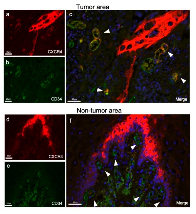
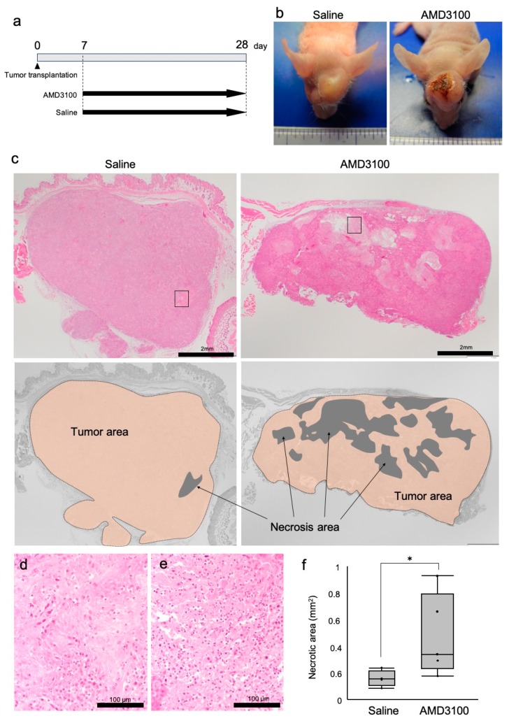
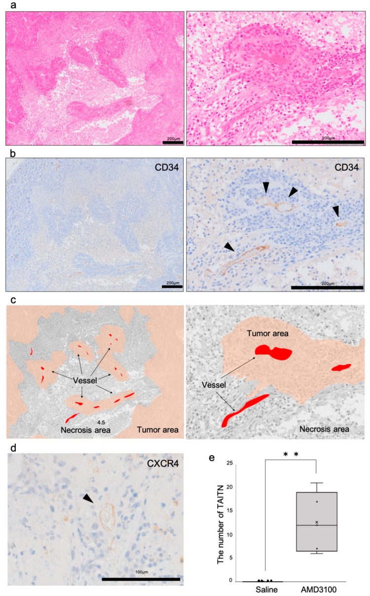
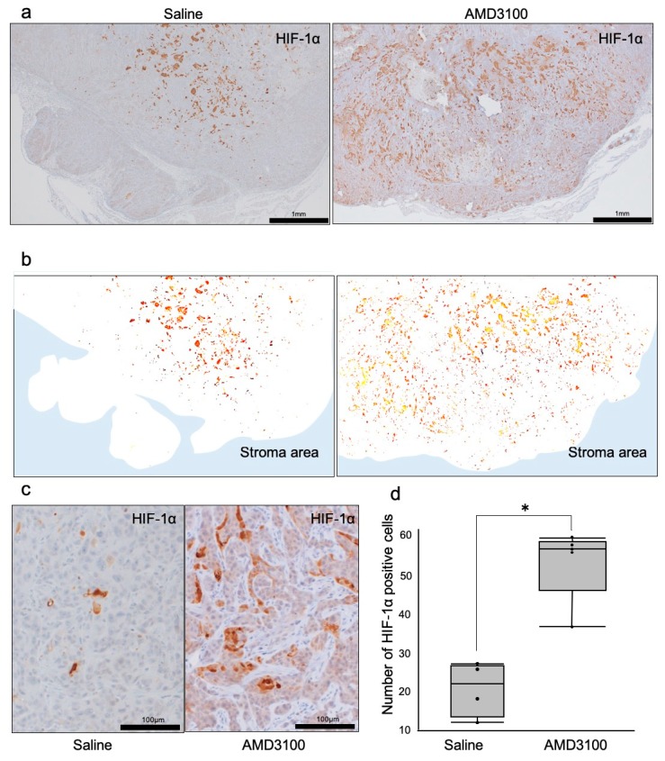
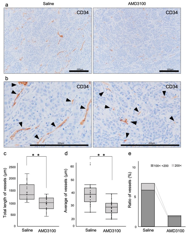
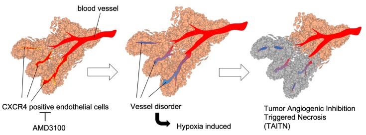
CXCR4 is a chemokine receptor crucial in tumor progression, although the angiogenic role of CXCR4 in oral squamous cell carcinoma (OSCC) has not been investigated. Here we show that CXCR4 is crucial for tumor angiogenesis, thereby supporting tumor survival in OSCC. Immunohistochemistry on human clinical specimens revealed that CXCR4 and a tumor vasculature marker CD34 were co-distributed in tumor vessels in human OSCC specimens. To uncover the effects of CXCR4 inhibition, we treated the OSCC-xenografted mice with AMD3100, so-called plerixafor, an antagonist of CXCR4. Notably, we found a unique pathophysiological structure defined as tumor angiogenic inhibition triggered necrosis (TAITN), which was induced by the CXCR4 antagonism. Treatment with AMD3100 increased necrotic areas with the induction of hypoxia-inducible factor-1α in the xenografted tumors, suggesting that AMD3100-induced TAITN was involved in hypoxia and ischemia. Taken together, we demonstrated that CXCR4 plays a crucial role in tumor angiogenesis required for OSCC progression, whereas TAITN induced by CXCR4 antagonism could be an effective anti-angiogenic therapeutic strategy in OSCC treatment.

Keywords: CXCR4 antagonist; hypoxia; oral squamous cell carcinoma; tumor angiogenic inhibition triggered necrosis (TAITN); tumor blood vessel.


Conflict of interest statement

The authors declare no conflict of interest.

Figures

Figure 1
Figure 1
Investigation of CXCR4-positive and CD34-positive vessels in oral squamous cell carcinoma (OSCC) stroma. (a) HE staining of an OSCC tissue for the definition of the tumor and nontumor areas. Tumor and nontumor areas are surrounded by dotted lines. (b) High-power magnification of tumor area stained with HE. Tu: tumor. St: stroma. (c) Immunohistochemistry (IHC) for CD34 in tumor and nontumor areas. Borders between epithelia (Ep), connective tissue (Co), tumor (Tu), and stroma (St) are shown with dotted lines. (d) The average number of vessels in the tumor and nontumor areas in a representative OSCC case. p = 0.289, n.s., not significant, N = 10 cases. (e) IHC for CXCR4 in tumor and nontumor areas. (f) High magnification IHC for CXCR4. Arrowheads indicate vessels. CXCR4-positive vessels specifically existed in the tumor area. (g) The average number of CXCR4-positive vessels in the tumor and nontumor areas in a representative OSCC case. ** p < 0.0001, N = 10 cases.
Figure 2
Figure 2
Double-fluorescent IHC for CXCR4 and CD34 in tumor and nontumor areas. (a) CXCR4 stained in stromal vessels and tumor cells (red). (b) CD34 stained only on vessels in the tumor stroma (green). (c) A merged IHC image of CD34 and CXCR4. Nuclei were stained with DAPI. Arrowheads indicate CXCR4/CD34 double-positive tumor vessels in the OSCC stroma. (d) CXCR4 stained in the nontumor area (red). (e) CD34 stained in the nontumor area (green). (f) A merged IHC image of CD34 and CXCR4. Nuclei were stained with DAPI. CD34-positive endothelial cells were all negative for CXCR4 (arrowheads).
Figure 3
Figure 3
CXCR4 antagonist AMD3100 induced tumor necrosis. Mice were injected with HSC-2 cells subcutaneously in the back and head. Seven days after tumor transplantation, AMD3100 or saline was intraperitoneally administrated every day for 21 d. (a) A schematic protocol of tumor injection and AMD3100 administration. (b) Representative photographs of subcutaneous tumors formed in mice. Necrosis and bleeding were observed only in the AMD3100 group. (c) Definition of necrotic areas. Top, HE staining. The areas closed with rectangles are enlarged in “d” and “e”. Bottom, diagrams defining necrotic areas in each tumor. (d,e) High-power magnification views of tumor necrotic areas in the saline-treated mouse (d) and AMD3100-treated mouse (e). The areas are enlarged from Figure 3c. (f) Box-whisker-dot plot of tumor necrotic areas with or without AMD3100 administration. * p = 0.0416, N = 5 mice.
Figure 4
Figure 4
Pathological analysis of tumor angiogenic inhibition triggered necrosis (TAITN) induced by CXCR4 antagonism. (a) Representative HE staining of tumor necrotic area in an AMD3100-treated mouse. (b) Representative IHC for CD34 in the tumor necrotic area in an AMD3100-treated mouse. (c) Illustration of the structure of TAITN found in AMD3100-treated mice. Gray, necrotic area. Red, tumor vessels. Brown, tumor area. Islanded tumor areas are surrounded by necrotic area. The center of each tumor island is occupied by vessels. We defined this unique pathophysiological conception as TAITN. (d) IHC for CXCR4 in the TAITN. CXCR4-positive blood vessels were observed at the center of TAITN. (e) The number of vessel-tumor islands in the AMD3100 group and saline group. ** p = 0.0031, N = 5 mice.
Figure 5
Figure 5
Distribution of hypoxia-inducible factor-1α (HIF-1α) in tumors treated with or without CXCR4 antagonist AMD3100. (a) IHC for HIF-1α in the tumor area under low-power magnification. (b) Illustration of HIF-1α distribution in tumors. White area, tumor. Blue area, stroma. (c) IHC of HIF-1α under high-power magnification. (d) The number of HIF-1α-positive tumor cells. * p = 0.02, N = 5 mice.
Figure 6
Figure 6
Tumor vessels were disorganized by CXCR4 antagonism. (a) Low-power magnification IHC of CD34. (b) High-power magnification IHC of CD34. (c) The total length of CD34-positive vessels. ** p < 0.0001, N = 25. (d) The average length of CD34-positive vessels. ** p < 0.0001, N = 25. (e) Rate of vessels sized 100~200 μm, and >200 μm.
Figure 7
Figure 7
Graphical abstract: CXCR4 antagonism induced tumor necrosis through vessel disorder. Left, a CXCR4 antagonist AMD3100 inhibited CXCR4-positive endothelial cells of tumor vessels. Middle, inhibition of CXCR4-positive endothelial cells induced vessel disorder and caused hypoxia in surrounding tumor cells. Right, necrosis was caused in the surrounding tumor cells through vessel disorder. Taken together, we propose a novel mechanistic concept of “Tumor Angiogenic Inhibition Triggered Necrosis (TAITN)”.

Similar articles

Cited by

References

    1. Bleul C.C., Wu L., Hoxie J.A., Springer T.A., Mackay C.R. The HIV coreceptors CXCR4 and CCR5 are differentially expressed and regulated on human T lymphocytes. Proc. Natl. Acad. Sci. USA. 1997;94:1925–1930. doi: 10.1073/pnas.94.5.1925. - DOI - PMC - PubMed
    1. Bleul C.C., Farzan M., Choe H., Parolin C., Clark-Lewis I., Sodroski J., Springer T.A. The lymphocyte chemoattractant SDF-1 is a ligand for LESTR/fusin and blocks HIV-1 entry. Nature. 1996;382:829–833. doi: 10.1038/382829a0. - DOI - PubMed
    1. Hauser A.E., Debes G.F., Arce S., Cassese G., Hamann A., Radbruch A., Manz R.A. Chemotactic Responsiveness Toward Ligands for CXCR3 and CXCR4 Is Regulated on Plasma Blasts During the Time Course of a Memory Immune Response. J. Immunol. 2002;169:1277–1282. doi: 10.4049/jimmunol.169.3.1277. - DOI - PubMed
    1. Nakayama T., Hieshima K., Izawa D., Tatsumi Y., Kanamaru A., Yoshie O. Cutting edge: Profile of chemokine receptor expression on human plasma cells accounts for their efficient recruitment to target tissues. J. Immunol. 2003;170:1136–1140. doi: 10.4049/jimmunol.170.3.1136. - DOI - PubMed
    1. Schecter A.D., Berman A.B., Taubman M.B. Chemokine receptors in vascular smooth muscle. Microcirculation. 2003;10:265–272. doi: 10.1080/mic.10.3-4.265.272. - DOI - PubMed

Publication types

MeSH terms