. 2019 Jul 22;11(7):1034.
doi: 10.3390/cancers11071034.

Flavopereirine Suppresses the Growth of Colorectal Cancer Cells through P53 Signaling Dependence

Affiliations

Flavopereirine Suppresses the Growth of Colorectal Cancer Cells through P53 Signaling Dependence

Jhy-Ming Li et al. Cancers (Basel). .

Abstract

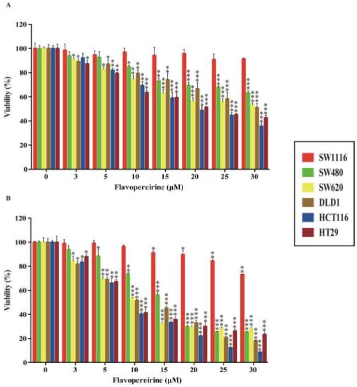
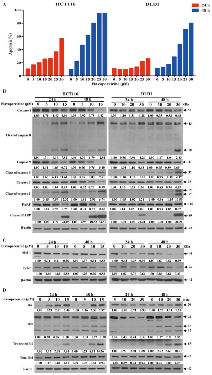
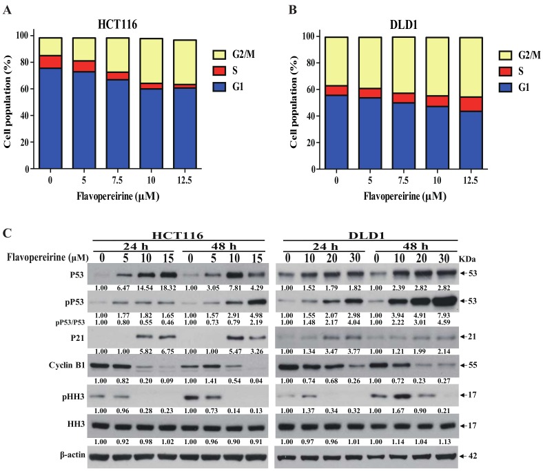
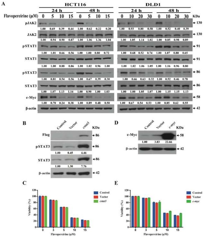
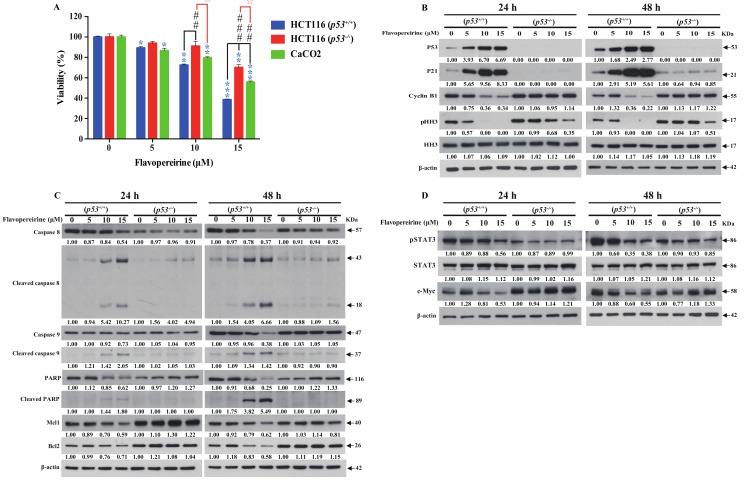
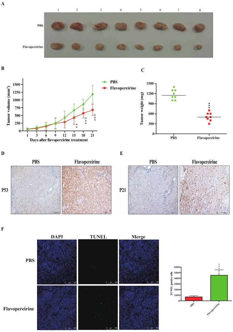
Colorectal cancer (CRC) is a significant cause of morbidity and mortality worldwide. The outcome of CRC patients remains poor. Thus, a new strategy for CRC treatment is urgently needed. Flavopereirine is a β-carboline alkaloid extracted from Geissospermum vellosii, which can reduce the viability of various cancer cells through an unknown mode of action. The aim of the present study was to investigate the functional mechanism and therapeutic potential of flavopereirine on CRC cells in vitro and in vivo. Our data showed that flavopereirine significantly lowered cellular viability, caused intrinsic and extrinsic apoptosis, and induced G2/M-phase cell cycle arrest in CRC cells. Flavopereirine downregulated Janus kinases-signal transducers and activators of transcription (JAKs-STATs) and cellular myelocytomatosis (c-Myc) signaling in CRC cells. In contrast, the enforced expressions of constitutive active STAT3 and c-Myc could not restore flavopereirine-induced viability reduction. Moreover, flavopereirine enhanced P53 expression and phosphorylation in CRC cells. CRC cells with P53 knockout or loss-of-function mutation significantly diminished flavopereirine-mediated viability reduction, indicating that P53 activity plays a major role in flavopereirine-mediated CRC cell growth suppression. Flavopereirine also significantly repressed CRC cell xenograft growth in vivo by upregulating P53 and P21 and inducing apoptosis. In conclusion, flavopereirine-mediated growth suppression in CRC cells depended on the P53-P21, but not the JAKs-STATs-c-Myc signaling pathway. The present study suggests that flavopereirine may be efficacious in the clinical treatment of CRC harboring functional P53 signaling.

Keywords: Flavopereirine; P53; colorectal cancer.


Conflict of interest statement

The authors declare no conflicts of interest.

Figures

Figure 1
Figure 1
Flavopereirine lowered the viability of various CRC cell lines. CRC cells were treated with various concentrations of flavopereirine for 24 h and 48 h and their viability was assayed by CCK8. Values were expressed as cell viability [%]. Each value represented the mean ± SD of quadruplicate assays. * p < 0.05; ** p < 0.01, *** p < 0.001 compared with the control. Similar results were obtained for at least three independent experiments.
Figure 2
Figure 2
Flavopereirine induced both extrinsic and intrinsic apoptosis in CRC cells. (A) Flavopereirine-induced apoptosis in HCT116 and DLD1 cells was assayed and quantified after 24 h and 48 h treatment. (B) Flavopereirine promoted the cleavage of caspase-8, -9, and -3 and PARP. (C,D) Flavopereirine downregulated the pro-survival Bcl-2 family proteins and upregulated the pro-apoptotic proteins Mcl-1, Bcl-2, Bik, Bim, and Truncated Bid. Numbers under the plots indicate the fold change of protein level relative to their untreated control. Similar results were obtained in at least three independent experiments.
Figure 3
Figure 3
Flavopereirine induced G2/M-phase cell cycle arrest. (A) The cell cycle distributions of HCT116 and DLD1 were assayed and quantified after flavopereirine treatment for 24 h. Values were expressed as cell population [%]. (B) Flavopereirine upregulated P53 and P21 expression and phosphorylation, downregulated cyclin B1, and diminished the phosphorylation of histone H3 after 24 h and 48 h treatment. Numbers under the plots indicate the fold change of protein level relative to their untreated control. Similar results were obtained for at least three independent experiments.
Figure 4
Figure 4
Flavopereirine non-dependently utilized STAT3/c-Myc signaling to reduce CRC cell viability. (A) Phosphorylation of JAK2, STAT1 and STAT3 and expression of downstream c-Myc protein were suppressed in a dose-dependent manner after treatment with flavopereirine for 24 h and 48 h. (B,D) STAT3 and c-Myc proteins were strongly upregulated by introducing cstat3 and c-myc into CRC cells at 48 h. (C,E) The enforced expressions of STAT3 and c-Myc did not reverse flavopereirine-mediated viability reduction in CRC cells. Numbers under the plots indicate the fold change of protein level relative to their untreated control. Similar results were obtained for at least three independent experiments.
Figure 5
Figure 5
P53 signaling participated in flavopereirine-mediated viability reduction and apoptosis induction. HCT116 p53+/+, p53−/− and CaCO2 cells were treated with flavopereirine for 48 h. (A) Cell viability was determined by CCK8 assay. Each value was the mean ± SD of quadruplicate assays. * p < 0.05; ** p < 0.01, *** p < 0.001 compared to each untreated control. ## p < 0.01 and ### p < 0.001 compared to flavopereirine (10 or 15 µM) treated HCT116 (p53+/+) group. p < 0.05 and ☆☆p < 0.01 compared to flavopereirine (10 or 15 µM) treated HCT116 (p53−/−) group. (BD) Pro-apoptotic, anti-survival, G2/M-phase cell cycle, and STAT3 signaling were not changed in HCT116 (p53−/−) cells relative to wild type HCT116 (p53+/+) cells after flavopereirine treatment for 24 h and 48 h. Numbers under the plots indicate the fold change of protein level relative to their untreated control. Similar results were obtained in at least three independent experiments.
Figure 6
Figure 6
Flavopereirine significantly inhibited in vivo HCT116-xenograft growth. HCT116-bearing mice were treated either with PBS (n = 8) or flavopereirine (10 mg kg−1 d−1) (n = 8). (A) Representative images of excised tumors from each group. (B) Tumor volumes were measured to reflect tumor growth. ** p < 0.01, *** p < 0.001 compared with PBS. (C) Tumor weights were compared on the last day of treatment. *** p < 0.001 compared with PBS. (D,E) P53 and P21 expression levels in HCT116 tumors were evaluated by immunohistochemical staining. Scale bar: 100 µm. (F) Apoptosis in HCT116 tumors was measured by TUNEL staining. *** p < 0.001 compared with PBS. Scale bar: 100 µm.

Similar articles

Cited by

References

    1. Favoriti P., Carbone G., Greco M., Pirozzi F., Pirozzi R.E., Corcione F. Worldwide burden of colorectal cancer: A review. Updates Surg. 2016;68:7–11. doi: 10.1007/s13304-016-0359-y. - DOI - PubMed
    1. Karapetis C.S., Khambata-Ford S., Jonker D.J., O’Callaghan C.J., Tu D., Tebbutt N.C., Simes R.J., Chalchal H., Shapiro J.D., Robitaille S., et al. K-ras mutations and benefit from cetuximab in advanced colorectal cancer. N. Engl. J. Med. 2008;359:1757–1765. doi: 10.1056/NEJMoa0804385. - DOI - PubMed
    1. Sun X., Wang X., Feng W., Guo H., Tang C., Lu Y., Xiang X., Bao Y. Gene signatures associated with drug resistance to irinotecan and oxaliplatin predict a poor prognosis in patients with colorectal cancer. Oncol. Lett. 2017;13:2089–2096. doi: 10.3892/ol.2017.5691. - DOI - PMC - PubMed
    1. Bardhan K., Liu K. Epigenetics and colorectal cancer pathogenesis. Cancers. 2013;5:676–713. doi: 10.3390/cancers5020676. - DOI - PMC - PubMed
    1. Brenner H., Kloor M., Pox C.P. Colorectal cancer. Lancet. 2014;383:1490–1502. doi: 10.1016/S0140-6736(13)61649-9. - DOI - PubMed