. 2019 Jul 13;11(7):407.
doi: 10.3390/toxins11070407.

Development and Characterization of Monoclonal Antibodies to Botulinum Neurotoxin Type E

Affiliations

Development and Characterization of Monoclonal Antibodies to Botulinum Neurotoxin Type E

Candace S Bever et al. Toxins (Basel). .

Abstract

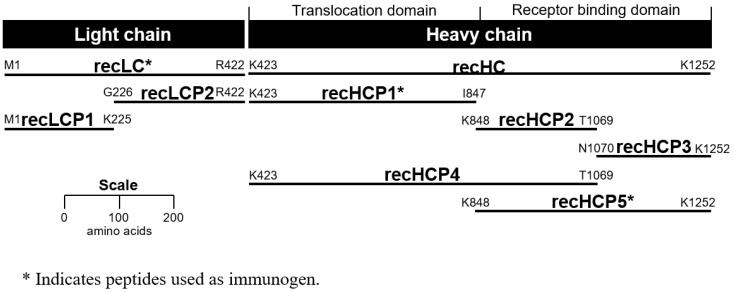
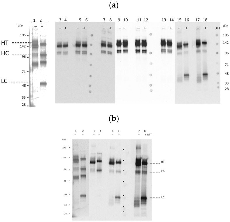
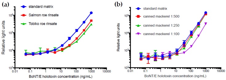
Botulism is a devastating disease caused by botulinum neurotoxins (BoNTs) secreted primarily by Clostridium botulinum. Mouse bioassays without co-inoculation with antibodies are the standard method for the detection of BoNTs, but are not capable of distinguishing between the different serotypes (A-G). Most foodborne intoxications are caused by serotypes BoNT/A and BoNT/B. BoNT/E outbreaks are most often observed in northern coastal regions and are associated with eating contaminated marine animals and other fishery products. Sandwich enzyme-linked immunosorbent assays (ELISAs) were developed for the detection of BoNT/E3. Monoclonal antibodies (mAbs) were generated against BoNT/E3 by immunizing with recombinant peptide fragments of the light and heavy chains of BoNT/E3. In all, 12 mAbs where characterized for binding to both the recombinant peptides and holotoxin, as well as their performance in Western blots and sandwich ELISAs. The most sensitive sandwich assay, using different mAbs for capture and detection, exhibited a limit of detection of 0.2 ng/ml in standard buffer matrix and 10 ng/mL in fish product matrices. By employing two different mAbs for capture and detection, a more standardized sandwich assay was constructed. Development of sensitive and selective mAbs to BoNT/E would help in the initial screening of potential food contamination, speeding diagnosis and reducing use of laboratory animals.

Keywords: BoNT/E; ELISA; botulinum neurotoxin serotype E; immunoassay; monoclonal antibodies.


Conflict of interest statement

The authors declare no conflicts of interest. The funders had no role in the design of the study; in the collection, analyses, or interpretation of data; in the writing of the manuscript, or in the decision to publish the results.

Figures

Figure 1
Figure 1
Peptide fragments of botulinum neurotoxins (BoNT)/E3. Glutathione S-transferase (GST) was fused on the N-terminus of each peptide.
Figure 2
Figure 2
Western blots of BoNT/E3 nicked holotoxin with and without dithiothreitol (DTT). Bands (HT = holotoxin; HC = heavy chain; LC = light chain) were visualized by the following: (a) lanes 1–2, silver stain; lanes 3–4, BoE 71-9; lanes 5–6, BoE 71-14; lanes 7–8, BoE 71-95; lanes 9–10, BoE 66-29; lanes 11–12, BoE 66-81; lanes 13–14, BoE 66-106; lanes 15–16, BoE 33-13-17-4; lanes 17–18, BoE 34-16; (b) lanes 1–2, silver stain; lanes 3–4 BoE 9-15; lanes 5–6, BoE 33-10; 7–8 BoE 33-12. (BoE 33-17-9 not shown). The silver stained (lanes 1–2) band ~80 kDa is unknown/unidentified material not recognized by the antibodies.
Figure 3
Figure 3
Epitope identification of anti-BoNT/E3 mAbs. Epitope location is based on antibody binding to recombinant toxin-GST fusion peptides. Diagram is drawn to approximate scale. The predicted transmembrane (K423 to I847) and receptor binding (K848 to K1253) domains are indicated.
Figure 4
Figure 4
A capture ELISA for the detection of BoNT/E3 holotoxin using mAb BoE 66-29 as the capture antibody paired with biotinylated mAbs BoE 9-15, BoE 71-9, or BoE 71-95 as the detector antibody. Points represent average (n = 3) measurements, error bars are standard deviation, and the horizontal dashed lines equal the average of the zero spike plus three standard deviation (SD). Where not visible, error bars are within the symbols.
Figure 5
Figure 5
Analysis of matrix effects in the BoNT/E3 holotxin sandwich ELISA. The matrices (a) Salmon and Tobiko roe rinsates and (b) canned mackerel at dilutions of 1:100, 1:250, and 1:500 were spiked with BoNT/E3 holotoxin and are shown alongside the standard curve (blue circles) using 3% non-fat dried milk in tris buffered saline with Tween 20 (TBST) as the matrix. Where not visible, error bars are within the symbols.

References

    1. Peck M.W., Smith T.J., Anniballi F., Austin J.W., Bano L., Bradshaw M., Cuervo P., Cheng L.W., Derman Y., Dorner B.G., et al. Historical perspectives and guidelines for botulinum neurotoxin subtype nomenclature. Toxins. 2017;9:38. doi: 10.3390/toxins9010038. - DOI - PMC - PubMed
    1. Zhang S., Masuyer G., Zhang J., Shen Y., Lundin D., Henriksson L., Miyashita S.I., Martinez-Carranza M., Dong M., Stenmark P. Identification and characterization of a novel botulinum neurotoxin. Nat. Commun. 2017;8:14130. doi: 10.1038/ncomms14130. - DOI - PMC - PubMed
    1. CDC . Botulism Annual Summary, 2016. US Department of Health and Human Services; Atlanta, Georgia: 2017. [(accessed on 1 April 2019)]. Available online: https://www.cdc.gov/botulism/pdf/Botulism-2016-SUMMARY-508.pdf.
    1. Carter A.T., Peck M.W. Genomes, neurotoxins and biology of Clostridium botulinum group I and group II. Res. Microbiol. 2015;166:303–317. doi: 10.1016/j.resmic.2014.10.010. - DOI - PMC - PubMed
    1. Hyytia E., Hielm S., Korkeala H. Prevalence of Clostridium botulinum type E in finnish fish and fishery products. Epidemiol. Infect. 1998;120:245–250. doi: 10.1017/S0950268898008693. - DOI - PMC - PubMed

Publication types

MeSH terms

LinkOut - more resources