Review
. 2019 Jul 14;9(7):279.
doi: 10.3390/biom9070279.

Flagella-Driven Motility of Bacteria

Affiliations
Review

Flagella-Driven Motility of Bacteria

Shuichi Nakamura et al. Biomolecules. .

Abstract

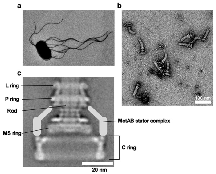
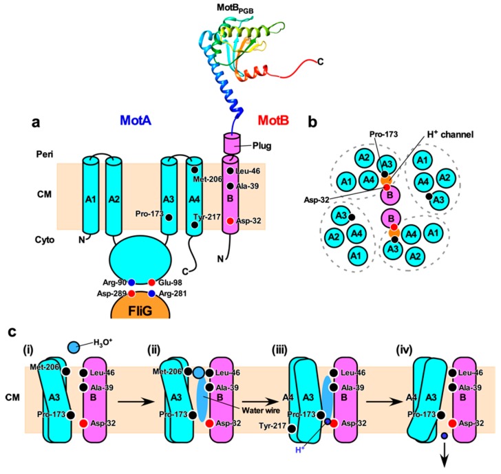
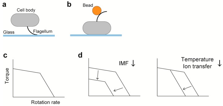
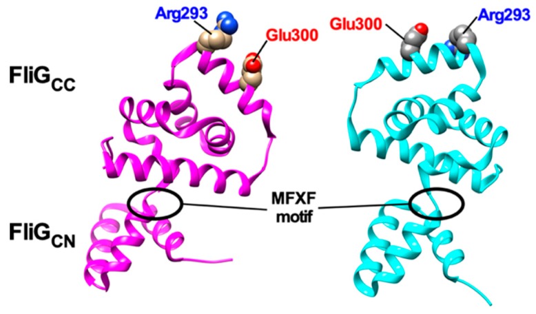
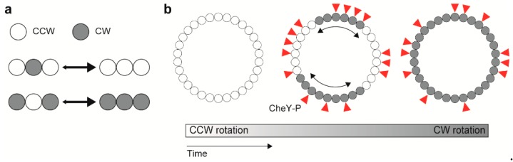
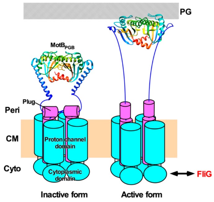
The bacterial flagellum is a helical filamentous organelle responsible for motility. In bacterial species possessing flagella at the cell exterior, the long helical flagellar filament acts as a molecular screw to generate thrust. Meanwhile, the flagella of spirochetes reside within the periplasmic space and not only act as a cytoskeleton to determine the helicity of the cell body, but also rotate or undulate the helical cell body for propulsion. Despite structural diversity of the flagella among bacterial species, flagellated bacteria share a common rotary nanomachine, namely the flagellar motor, which is located at the base of the filament. The flagellar motor is composed of a rotor ring complex and multiple transmembrane stator units and converts the ion flux through an ion channel of each stator unit into the mechanical work required for motor rotation. Intracellular chemotactic signaling pathways regulate the direction of flagella-driven motility in response to changes in the environments, allowing bacteria to migrate towards more desirable environments for their survival. Recent experimental and theoretical studies have been deepening our understanding of the molecular mechanisms of the flagellar motor. In this review article, we describe the current understanding of the structure and dynamics of the bacterial flagellum.

Keywords: bacterial flagellum; chemotaxis; ion channel; ion motive force; mechanochemical coupling; molecular motor; motility; torque generation.


Conflict of interest statement

The authors declare no conflict of interest.

Figures

Figure 1
Figure 1
Salmonella flagellum. (a) Electron micrograph of Salmonella cell. The micrograph was taken at a magnification of ×1200. (b) Electron micrograph of hook-basal bodies isolated from Salmonella cells. (c) CryoEM image of purified basal body. Purified basal body consists of the L, P, MS and C rings and the rod. A dozen MotAB complex are associated with the basal body to act as a stator unit in the motor but is gone during purification.
Figure 2
Figure 2
H+ translocation mechanism of the flagellar motor. (a) Topology of the E. coli MotA and MotB and a crystal structure of the peptidoglycan-binding domain of MotB (MotBPGB, PDB code: 2ZVY). Highly conserved Arg-90 and Glu-98 residues in the cytoplasmic loop between transmembrane helices 2 (A2) and 3 (A3) interact with conserved Asp-289 and Arg-281 residues of FliG, respectively, to drive motor rotation. Asp-32 of MotB provides a binding site for H+. Pro-173, Met-206 and Tyr-217 of MotA and Ala-39 and Leu-46 of MotB are involved in the H+ relay mechanism. Cyto, cytoplasm; CM, cytoplasmic membrane; Peri, periplasm. (b) Arrangement of transmembrane segments of MotA and MotB. The MotAB complex has two proton channels. Four MotA subunits are positioned with their TM3 (A3) and TM4 (A4) segments adjacent to the MotB dimer, and their TM1 (A1) and TM2 (A2) segments on the outside. (c) A plausible model for H+ translocation through MotAB stator complex (see text for details).
Figure 3
Figure 3
Characterization of the rotation of the flagellar motor. (a) Tethered cell assay. (b) Bead assay; gold nanoparticles (60–100 nm in diameter) and polystyrene beads (0.2–2.0 μm in diameter) are used. (c) A schematic of the torque-speed curve. (d) Effects of factors relevant to motor dynamics on the torque-speed curve. Dependence of the curve on the number of stator units is described in the section of Duty ratio.
Figure 4
Figure 4
Structural comparisons between 3USY (cyan) and 3USW (magenta) structures of Helicobacter pylori FliG. Conformational rearrangements of the conserved MFXF motif induces a 180° rotation of FliGCC relative to FliGCN to reorient Arg-293 and Glu-300 residues, which correspond to Arg-281 and Asp-289 of E. coli FliG, respectively.
Figure 5
Figure 5
Model for cooperative switching between counterclockwise (CCW) and clockwise (CW) rotations. (a) Interaction between adjacent rotor subunits. (b) Conformational spread upon CheY-P binding.
Figure 6
Figure 6
Activation mechanism of the H+-type MotAB complex. The MotAB complex consists of at least three structural parts: a cytoplasmic domain, a transmembrane ion channel and a peptidoglycan-binding domain [MotBPGB, PDB codes: 2ZVY (left panel) and 5Y40 (right panel). When the MotAB complex adopts a compact conformation, a plug segment of MotB binds to a transmembrane H+ channel to suppress massive H+ flow (left). When the MotAB complex encounters a rotor, electrostatic interactions between the cytoplasmic domain of MotA and FliG trigger the dissociation of the plug segment from the channel, followed by partial unfolding of the N-terminal portion of MotBPGB to allow MotBPGB to bind to the peptidoglycan (PG) layer. As a result, the MotAB complex becomes an active H+-type stator unit to drive flagellar motor rotation (right).

Similar articles

Cited by

References

    1. Josenhans C., Suerbaum S. The role of motility as a virulence factor in bacteria. Int. J. Med. Microbiol. 2002;291:605–614. doi: 10.1078/1438-4221-00173. - DOI - PubMed
    1. Lertsethtakarn P., Ottemann K.M., Hendrixson D.R. Motility and chemotaxis in Campylobacter and Helicobacter. Annu. Rev. Microbiol. 2011;65:389–410. doi: 10.1146/annurev-micro-090110-102908. - DOI - PMC - PubMed
    1. Visick K.L. An intricate network of regulators controls biofilm formation and colonization by Vibrio fischeri. Mol. Microbiol. 2009;74:782–789. doi: 10.1111/j.1365-2958.2009.06899.x. - DOI - PMC - PubMed
    1. Scharf B.E., Hynes M.F., Alexandre G.M. Chemotaxis signaling systems in model beneficial plant–bacteria associations. Plant Mol. Biol. 2016;90:549–559. doi: 10.1007/s11103-016-0432-4. - DOI - PubMed
    1. Ishikawa T., Yoshida N., Ueno H., Wiedeman M., Imai Y., Yamaguchi T. Energy transport in a concentrated suspension of bacteria. Phys. Rev. Lett. 2011;107:028102. doi: 10.1103/PhysRevLett.107.028102. - DOI - PubMed

Publication types

MeSH terms

Substances

LinkOut - more resources