. 2019 Jul 14;11(7):982.
doi: 10.3390/cancers11070982.

Docosahexaenoic Acid Enhances Oxaliplatin-Induced Autophagic Cell Death via the ER Stress/Sesn2 Pathway in Colorectal Cancer

Affiliations

Docosahexaenoic Acid Enhances Oxaliplatin-Induced Autophagic Cell Death via the ER Stress/Sesn2 Pathway in Colorectal Cancer

Soyeon Jeong et al. Cancers (Basel). .

Abstract

Oxaliplatin is an anticancer drug administered to colorectal cancer (CRC) patients in combination with 5-fluorouracil and antibodies (bevacizumab and cetuximab), thereby significantly improving the survival rate of CRC. However, due to various side effects associated with the above treatment strategy, the need for combinatorial therapeutic strategies has emerged. Based on the demand for new combinatorial therapies and the known antitumor effects of the omega-3 polyunsaturated fatty acid, docosahexaenoic acid (DHA), we investigated the Oxaliplatin and DHA combination for its effect. Our results indicated that DHA further enhanced Oxaliplatin-induced cell viability and autophagic cell death, in vitro and in vivo. Oxaliplatin and DHA also increased the expression of Sestrin 2 (SESN2) and endoplasmic reticulum (ER) stress related C/EBP homologous protein (CHOP). Additionally, treatment with Oxaliplatin and DHA enhanced the binding of CHOP to the promotor region of SESN2, increasing SESN2 expression. These results suggested that DHA enhanced Oxaliplatin-induced reduction in cell viability and increase in autophagy via activating SESN2 and increasing ER stress. Thus, SESN2 may be an effective preclinical target for CRC treatment.

Keywords: Oxaliplatin; Sestrin 2; autophagic cell death; colon cancer; docosahexaenoic acid.


Conflict of interest statement

The authors declare that they have no competing interests.

Figures

Figure 1
Figure 1
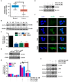
Combinatorial treatment with Oxaliplatin and docosahexaenoic acid (DHA) reduces viability and induces cell death in human colorectal cancer (CRC) cells. (A) Human normal colon CCD-18Co cells and various human CRC cells were treated with 0–60 µM of DHA for 24 h. Cell viability was measured via a WST-1 assay. ***, p < 0.001; (B) HCT116 cells were treated with 0–60 µM of ω3-polyunsaturated fatty acids (PUFAs) or ω6-PUFAs for 24 h. Cell viability was determined via a WST-1 assay; (C) Combination index (CI) for Oxaliplatin and DHA; (D) CRC cells were treated with the indicated doses of Oxaliplatin and DHA for 24 h. Cell viability was determined via the WST-1 assay. ***, p < 0.001; (E) Cell death was measured via Annexin V/propidium iodide staining using flow cytometry in HCT116 cells. ***, p < 0.001; (F) Intensity of bioluminescence after treatment with Oxaliplatin and DHA in the HCT116 Luc+ cells. The captured images were quantified using Image J. ***, p < 0.001.
Figure 2
Figure 2
DHA enhances Oxaliplatin-induced autophagy in human CRC cells. (A,B) HCT116 cells were subjected to indicated doses (A) and time (B) periods for 24 h. Then, the protein level of microtubule-associated protein 1A/1B-light chain 3 (LC3) and p62 were analyzed by immunoblotting. (C) Formation of green fluorescence protein (GFP)-LC3 puncta following exposure to Oxaliplatin and DHA was analyzed using confocal microscopy (Scale Bar, 10 μm). (D,E) HCT116 cells were treated with Oxaliplatin and DHA in the absence or presence of chloroquine (CQ) or rapamycin for 24, and 48 h. Autophagic cells (D), and autophagic markers (E) were detected using immunoblotting and a flow cytometer with an autophagy detection kit.
Figure 3
Figure 3
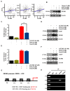
Combinatorial treatment upregulates expression of SESN2 in human CRC. (A) mRNA expression levels of SESN2 in normal and tumor tissues were evaluated using the mRNA expression data of the IlluminaHiSeq of TCGA CRC. **, p < 0.01. (B) SESN2 protein levels in normal and tumor tissues were evaluated using western blotting. (C,D) Comparison of endogenous expression levels of SESN2 mRNA and protein in normal human cells and human CRC cells using quantitative real-time polymerase chain reaction (qRT-PCR) analysis (C), immunoblotting (D). ***, p < 0.001. (E,F) SESN2 protein levels in the Oxaliplatin and DHA combination was analyzed by immunoblotting (E) and immunofluorescence (F) (Scale bar, 10 μm). (G,H) Following transfection with control small interfering RNA (siRNA) or SESN2 siRNA, cells were exposed to the Oxaliplatin and DHA combination. The mRNA (G) and protein (H) expression of LC3 and p62 were evaluated using qRT-PCR and western blotting. ***, p < 0.001.
Figure 4
Figure 4
Combined treatment with Oxaliplatin and DHA induces endoplasmic reticulum (ER) stress overproduction. (A,B) HCT116 cells were exposed to different doses of Oxaliplatin and DHA (A) and time periods (B). HCT116 cell lysates were analyzed with an immunoblotting assay using ER stress-related antibodies. (C) C/EBP homologous protein (CHOP) was confirmed via immunofluorescence in the Oxaliplatin and DHA combination (Scale Bar, 10 μM). (D,E) Following transfection with control siRNA or CHOP siRNA, the mRNA(D) and protein (E) levels of LC3 and p62 were detected via qRT-PCR and immunoblotting. **, p < 0.001. (F) Cells were exposed to Oxaliplatin and DHA with or without thapsigargin, and the p62 and LC3 levels were observed via western blotting.
Figure 5
Figure 5
CHOP regulates SESN2 activity by binding directly to the SESN2 promoter region. (A) The correlation of SESN2 expression and ER stress-related genes in CRC patients. (B) HCT116 cells were pre-treated with thapsigargin for 1 h and then exposed to the Oxaliplatin and DHA combination for 24 h. SESN2 protein expression was confirmed with western blotting. (CF) Following transfection with control siRNA, CHOP siRNA, or SESN2 siRNA, the SESN2 and CHOP protein levels were determined using qRT-PCR (C,E) and immunoblotting (D,F). ***, p < 0.001. (G) Illustration of the three predicted CHOP binding sites (BS) and ATF BS in the SESN2 promoter. (H) Cells were treated with Oxaliplatin and DHA, and then immunoprecipitated with either IgG or CHOP.
Figure 6
Figure 6
Combinatorial treatment with DHA and Oxaliplatin increases CHOP expression and in vivo autophagy. (AD) Nude mice were subcutaneously inoculated with 1 × 10⁷ HCT116-Luc+ Cells. When tumor volume reached approximately 200 mm3, the mice were treated with EtOH, DHA (10 mg/kg), Oxaliplatin (10 mg/kg), or the combinatorial treatment of DHA and Oxaliplatin (10 mg/kg), administered 3 times a week for 2 weeks. (A) Mice were imaged using the NightOWL LB983 bioluminescence imaging (BLI) system. (B) Line graph illustrating the tumor volume (mm3). ***, p < 0.001. (C) Tumor tissues were harvested on day 14 and then imaged using a digital camera. (D) Graph illustrating body weight. **, p < 0.01. (E,F) Immunohistochemistry (IHC) showing LC3 (upper panel), CHOP (middle panel), and SESN2 (lower panel) in the tumors from xenograft at a 400× magnification. (Scale bar, 20 μm). **, p < 0.01 and ***, p < 0.001.
Figure 7
Figure 7
Scheme of autophagic cell death induced by the combination of Oxaliplatin and DHA.

Similar articles

Cited by

References

    1. Siegel R.L., Miller K.D., Jemal A. Cancer statistics, 2018. CA Cancer J. Clin. 2018;68:7–30. doi: 10.3322/caac.21442. - DOI - PubMed
    1. Bendell J.C., Bekaii-Saab T.S., Cohn A.L., Hurwitz H.I., Kozloff M., Tezcan H., Roach N., Mun Y., Fish S., Flick E.D., et al. Treatment patterns and clinical outcomes in patients with metastatic colorectal cancer initially treated with FOLFOX-bevacizumab or FOLFIRI-bevacizumab: Results from ARIES, a bevacizumab observational cohort study. Oncologist. 2012;17:1486–1495. doi: 10.1634/theoncologist.2012-0190. - DOI - PMC - PubMed
    1. Mehmood R.K., Parker J., Ahmed S., Qasem E., Mohammed A.A., Zeeshan M., Jehangir E. Review of Cisplatin and Oxaliplatin in Current Immunogenic and Monoclonal Antibodies Perspective. World J. Oncol. 2014;5:97–108. doi: 10.14740/wjon830w. - DOI - PMC - PubMed
    1. Kim S.Y., Song X., Zhang L., Bartlett D.L., Lee Y.J. Role of Bcl-xL/Beclin-1 in interplay between apoptosis and autophagy in oxaliplatin and bortezomib-induced cell death. Biochem. Pharmacol. 2014;88:178–188. doi: 10.1016/j.bcp.2014.01.027. - DOI - PMC - PubMed
    1. Li D.D., Xie B., Wu X.J., Li J.J., Ding Y., Wen X.Z., Zhang X., Zhu S.G., Liu W., Zhang X.S., et al. Late-stage inhibition of autophagy enhances calreticulin surface exposure. Oncotarget. 2016;7:80842–80854. doi: 10.18632/oncotarget.13099. - DOI - PMC - PubMed