. 2019 Oct;33(10):11420-11430.
doi: 10.1096/fj.201900885R. Epub 2019 Jul 23.

SCF(FBXW7)-mediated degradation of p53 promotes cell recovery after UV-induced DNA damage

Affiliations

SCF(FBXW7)-mediated degradation of p53 promotes cell recovery after UV-induced DNA damage

María Galindo-Moreno et al. FASEB J. 2019 Oct.

Abstract

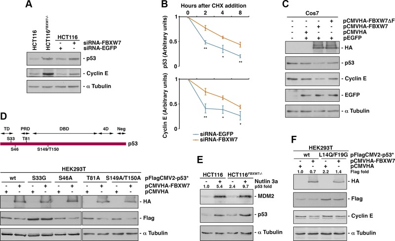
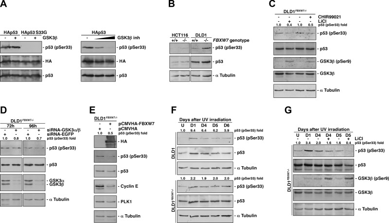
Eukaryotic cells have developed sophisticated mechanisms to ensure the integrity of the genome and prevent the transmission of altered genetic information to daughter cells. If this control system fails, accumulation of mutations would increase risk of diseases such as cancer. Ubiquitylation, an essential process for protein degradation and signal transduction, is critical for ensuring genome integrity as well as almost all cellular functions. Here, we investigated the role of the SKP1-Cullin-1-F-box protein (SCF)-[F-box and tryptophan-aspartic acid (WD) repeat domain containing 7 (FBXW7)] ubiquitin ligase in cell proliferation by searching for targets implicated in this process. We identified a hitherto-unknown FBXW7-interacting protein, p53, which is phosphorylated by glycogen synthase kinase 3 at serine 33 and then ubiquitylated by SCF(FBXW7) and degraded. This ubiquitylation is carried out in normally growing cells but primarily after DNA damage. Specifically, we found that SCF(FBXW7)-specific targeting of p53 is crucial for the recovery of cell proliferation after UV-induced DNA damage. Furthermore, we observed that amplification of FBXW7 in wild-type p53 tumors reduced the survival of patients with breast cancer. These results provide a rationale for using SCF(FBXW7) inhibitors in the treatment of this subset of tumors.-Galindo-Moreno, M., Giráldez, S., Limón-Mortés, M. C., Belmonte-Fernández, A., Reed, S. I., Sáez, C., Japón, M. Á., Tortolero, M., Romero, F. SCF(FBXW7)-mediated degradation of p53 promotes cell recovery after UV-induced DNA damage.

Keywords: FBXW7 inhibitors; cancer; proliferation; ubiquitylation.


Conflict of interest statement

This work was supported by Spanish grants from the Ministry of Economy and Competitiveness (MINECO; SAF2014-53799 and SAF2017-87358) and Junta de Andalucía (2017/BIO-211), and U.S. National Institutes of Health (NIH) National Cancer Institute Grant CA078343, and NIH National Institute of General Medical Sciences Grant GM115170 (to S.I.R.). A.B.-F. is the recipient of a Ph.D. fellowship from the Vicerrectorado de Investigación Plan Propio de Investigación (VI PPI) from Universidad de Sevilla. The authors declare no conflicts of interest.

Figures

Figure 1
Figure 1
Identification of p53 as a substrate of SCF(FBXW7). A) Amino acid sequence of p53 with matching peptides by mass spectrometry marked in yellow. B) Cos7 cells were transiently transfected with pFlagCMV2-FBXW7 and whole-cell extracts immunoprecipitated with anti-Flag mAb or normal mouse serum (PI). Immunoprecipitates materials were analyzed by Western blotting. Lys: whole-cell extracts from Cos7-transfected cells. C) The experiment was performed as in B, but proteins were immunoprecipitated with anti-p53 mAb or normal mouse serum. D) FBXW7 interacts with p53 in a yeast 2-hybrid assay. L40 reporter strain was cotransformed with the indicated constructs. The interaction between the 2-hybrid proteins is indicated by growth in the absence of histidine, yeast drop-out medium lacking Trp, Leu, and His (DO-wlh; dark red patch) and by the induction of LacZ expression (blue patches). RasV12/Raf interaction was used as a positive control. E) In vitro ubiquitylation assay of [35S]-labeled in vitro–transcribed and –translated p53 was conducted in the presence or absence of the following products: cold in vitro–transcribed and –translated FBXW7 or β-TrCP, E1 (His6-E1), E2 (His6-UbcH3 and UbcH5a), and ubiquitin (Ub). Samples were incubated at 30°C for 1 h. The bracket on the right side marks a ladder of bands corresponding to polyubiquitylated p53. F) The experiment was performed as in E, except that the unlabeled F-box protein was substituted by a recombinant SCF(FBXW7) complex expressed in Sf21 insect cells. G) Cos7 cells were transfected with the indicated plasmids and treated with MG132 for 4 h before harvesting. Extracts were prepared as indicated in the Materials and Methods and polyubiquitylated p53 visualized after Western blots of the Flag immunoprecipitations. AD, fusion with the activation domain of Gal4; BD, fusion with the DNA-binding domain of LexA; IP, immunoprecipitation.
Figure 2
Figure 2
SCF(FBXW7) induces p53 degradation in normal growing cells. A) Expression of p53 and cyclin E was studied in HCT116 and HCT116FBXW7−/− cells and in HCT116 cells transfected with the indicated siRNAs. An immunoblot for α-tubulin is shown as a loading control. B) U2OS cells were transfected with EGFP- or FBXW7-siRNA and, after 48 h, cycloheximide (CHX) was added to the medium, and cells were collected at the indicated times. Extracts were analyzed by Western blotting. p53 and cyclin E protein levels were quantified using ImageJ and normalized to α-tubulin levels. C) Cos7 cells were transfected with the indicated plasmids and, 18 h later, extracts were subjected to Western blot. D) Serines and threonines of the putative CPDs of p53 protein are indicated in the upper panel. In the lower panel, the overexpression of FBXW7 on WT or mutant p53 is shown. HEK293T cell were transfected with pFlagCMV2-p53 (WT or mutant) and 18 h later retransfected with pCMVHA-FBXW7 or empty vector. Extracts were analyzed by Western blot. E) HCT116 and HCT116FBXW7−/− cells were treated or not treated with Nutlin 3a for 24 h and extracts analyzed by immunoblot analysis. The quantitative fold change in p53 was determined relative to the loading control. F) Similarly to D, HEK293T were transfected with pFlagCMV2-p53 or pFlagCMV2-p53 L14Q/F19G and 18 h later retransfected with pCMVHA-FBXW7 or empty vector. Extracts were analyzed by Western blotting. The quantitative fold change in Flag-p53 or Flag-p53 L14Q/F19G was determined relative to the loading control. 4D, oligomerization domain; DBD, DNA-binding domain; Neg, negative regulation domain; PRD, proline-rich region; TD, transactivation domain. Error bars represent the sd (n = 3). *P < 0.05, **P < 0.01 (Student’s t test).
Figure 3
Figure 3
FBXW7 is involved in the recovery of cell proliferation after DNA damage. A) Cell proliferation assay of HCT116 and HCT116FBXW7−/− cells after UV radiation. The amount of protein in extracts was taken as a measure of cell proliferation. B) Extracts were also used to analyze p53 expression by Western blotting. C) Quantification of protein levels in B using ImageJ and normalized to α-tubulin level. D, days after UV irradiation. U, nonirradiated cells. Data are representative of 3 independent experiments. Error bars represent the sd (n = 4). *P < 0.05, **P < 0.01, ***P < 0.001 (Student’s t test).
Figure 4
Figure 4
GSK3 phosphorylates p53 on Ser33 and promotes its ubiquitylation by SCF(FBXW7) and degradation. A) In vitro kinase assay of cold in vitro–transcribed and –translated HAp53 and a p53 mutant on Ser33 (HAp53 S33G) incubated with recombinant GSK3-β. Proteins were analyzed by Western blotting using an anti–phosphorylated-p53 (Ser33) antibody (left panel). In the right panel, basal in vitro–transcribed and –translated HAp53 phosphorylation was inhibited with increasing amount of a specific GSK3-β inhibitor. B) Determination of phosphorylated p53 (Ser33) status of WT and FBXW7−/− HCT116 and DLD1 cells. C) Analysis of phosphorylated p53 (Ser33) status of DLD1FBXW7−/− cells treated or not treated with 2 GSK3 inhibitors (CHIR99021 and LiCl). D) DLD1FBXW7−/− cells were transfected with GSK3-α and -β or EGFP-siRNA, and 72 or 96 h later, cell extracts were blotted with the indicated antibodies. E) DLD1FBXW7−/− cells were transfected with pCMVHA-FBXW7 or pCMVHA, and extracts analyzed by Western blotting. F) Expression of phosphorylated p53 (Ser33) in DLD1 and DLD1FBXW7−/− cells after UV radiation. G) Effect of LiCl on p53 S33 phosphorylation of DLD1FBXW7−/− cells after UV irradiation. Inhibitor was added 24 h before harvesting. The quantitative fold change in phosphorylated p53 (Ser33) was determined relative to total p53 protein. Experiments were repeated 3 times with similar results. D, days after UV irradiation; U, untreated cells.
Figure 5
Figure 5
The role of FBXW7 in the recovery of cell proliferation after DNA damage is mediated by the degradation of p53. A) HCT116FBXW7−/− and HCT116p53−/− cells were transfected with the indicated plasmids and 24 h later irradiated (60 J/m2) or not. Crystal violet assay was performed from d 3 to 7 post-UV. Images show data from d 5 after irradiation. B) Quantification of crystal violet assay described in A. Error bars represent the sd (n = 3). The significance was obtained by comparing values of HCT116p53−/− pFlagCMV-p53 vs. HCT116p53−/− pFlagCMV-p53 S33G. Expression level of transfected plasmids is also shown. Scale bar, 400 μm. *P < 0.05, **P < 0.01, ***P < 0.001 (Student’s t test).
Figure 6
Figure 6
Influence of FBXW7 on apoptosis induced by p53 after DNA damage and its role in the survival of patients with breast cancer. A) HCT116 and DLD1 WT and FBXW7−/− cells were irradiated and collected at the indicated times. Extracts were analyzed by Western blotting. The quantitative fold change in cleaved PARP was determined relative to the loading control. Experiments were repeated 3 times with similar results. B) Kaplan-Meier analysis of patients with WT p53 breast cancer with WT FBXW7 (n = 1453; orange line) compared with increased levels of FBXW7, FBXW7amp (n = 68; blue line). Tick marks represent censored patients. The P values were obtained from the log-rank test of Mantel and Cox. C) Similar to B, but comparing patients with breast cancer with tumors p53loss/FBXW7wt (n = 852; orange line) vs. p53loss/FBXW7amp (n = 73; blue line). U, untreated control cells.

Similar articles

Cited by

References

    1. Ji C. H., Kwon Y. T. (2017) Crosstalk and interplay between the ubiquitin-proteasome system and autophagy. Mol. Cells 40, 441–449 - PMC - PubMed
    1. Kaiser P., Huang L. (2005) Global approaches to understanding ubiquitination. Genome Biol. 6, 233 - PMC - PubMed
    1. Nakayama K. I., Nakayama K. (2006) Ubiquitin ligases: cell-cycle control and cancer. Nat. Rev. Cancer 6, 369–381 - PubMed
    1. Welcker M., Orian A., Grim J. E., Eisenman R. N., Clurman B. E. (2004) A nucleolar isoform of the Fbw7 ubiquitin ligase regulates c-Myc and cell size. Curr. Biol. 14, 1852–1857; erratum: 15, 2285 - PubMed
    1. Grim J. E., Gustafson M. P., Hirata R. K., Hagar A. C., Swanger J., Welcker M., Hwang H. C., Ericsson J., Russell D. W., Clurman B. E. (2008) Isoform- and cell cycle-dependent substrate degradation by the Fbw7 ubiquitin ligase. J. Cell Biol. 181, 913–920 - PMC - PubMed

Publication types