Review
. 2019 Sep 29;6(1):93-117.
doi: 10.1146/annurev-virology-092917-043315. Epub 2019 Jul 23.

Physical and Functional Analysis of Viral RNA Genomes by SHAPE

Affiliations
Review

Physical and Functional Analysis of Viral RNA Genomes by SHAPE

Mark A Boerneke et al. Annu Rev Virol. .

Abstract

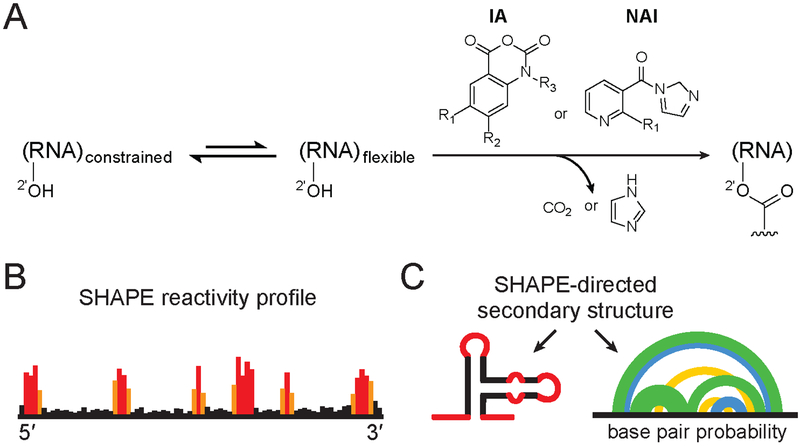
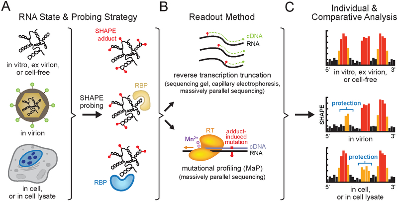
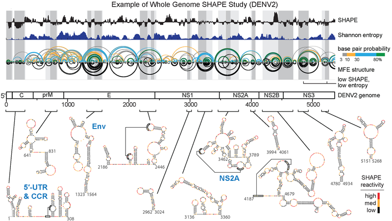
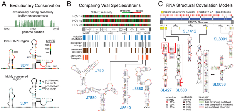
RNA viruses encode the information required to usurp cellular metabolism and gene regulation and to enable their own replication in two ways: in the linear sequence of their RNA genomes and in higher-order structures that form when the genomic RNA strand folds back on itself. Application of high-resolution SHAPE (selective 2'-hydroxyl acylation analyzed by primer extension) structure probing to viral RNA genomes has identified numerous new regulatory elements, defined new principles by which viral RNAs interact with the cellular host and evade host immune responses, and revealed relationships between virus evolution and RNA structure. This review summarizes our current understanding of genome structure-function interrelationships for RNA viruses, as informed by SHAPE structure probing, and outlines opportunities for future studies.

Keywords: RNA structure; RNA viruses; SHAPE; chemical probing; functional validation.


Figures

Figure 1
Figure 1
SHAPE chemistry mechanism, reagents used to probe viral RNA structure, and SHAPE-directed structure modeling. (A) SHAPE reagents react with the 2′-hydroxyl group of conformationally flexible RNA nucleotides, yielding a covalent 2’-O-adduct. Widely used reagents are based on the isatoic anhydride (IA) or nicotinic acid imidazolide (NAI) scaffolds (47, 54, 115). (B) Representative SHAPE reactivity profile, with black, orange and red bars indicating low, medium and high nucleotide reactivities. (C) Use of SHAPE data to model a single minimum free energy structure (left) or characterize an ensemble consistent with SHAPE data (right). Arcs (right) connect base paired nucleotides with pairing probabilities illustrated (from highest to lowest) in green, blue and yellow. Data in panels B and C correspond to the same representative structural element; adapted from Siegfried et al. (37).
Figure 2
Figure 2
SHAPE probing strategies. (A) Chemical probing of a viral RNA genome in an informative biological state (see Box 1 for definitions). Representative SHAPE adducts (red spheres) and RNA binding proteins (RBP) are shown. (B) Readout of per-nucleotide SHAPE reactivities by adduct-induced reverse transcription primer extension truncation or mutational profiling (MaP) and quantified by sequencing gel, capillary electrophoresis or massively parallel sequencing. (C) SHAPE reactivity profiles from distinct biological states can be compared to identify sites of SHAPE reactivity protections and enhancements, revealing state-specific RNA conformations, or protein or small molecule binding. Black, orange and red bars indicate low, medium and high nucleotide reactivities, respectively. Individual SHAPE reactivity profiles can also be incorporated into modeling algorithms to yield RNA secondary structure models (see Figure 1). Elements of figure adapted from Smola et al. (38, 116).
Figure 3
Figure 3
Whole viral genome studies and well-determined structures. Genome wide SHAPE structure probing from a representative study of the DENV2 RNA genome. The 5’ half of the ~11,000 nucleotide long RNA genome is shown. Median ex virion 1M7 SHAPE reactivities (black) and Shannon entropies (dark blue) are plotted over centered 55-nt windows. Regions with both low SHAPE and low Shannon entropy, termed low SHAPE, Shannon entropy regions, are highlighted by dark-gray shading, with light-gray shading extended to encompass entire structures. Base pairing arcs are colored by probability (see scale), with green arcs indicating the most probable and well-determined base pairs. The minimum free-energy (MFE) secondary structure (inverted black arcs) was obtained using SHAPE reactivities as restraints (37, 38, 117). Secondary structures of the twelve boxed low SHAPE, low Shannon entropy elements are shown at bottom of image and are colored by SHAPE reactivity. Previously studied RNA structural elements in the 5′-UTR and capsid-coding region (CCR) and elements correctly modeled de novo by SHAPE-directed modeling are labeled in blue. Novel structural elements in the Env- and NS2A- coding regions identified by SHAPE structure probing and shown to be critical for viral fitness are also labeled in blue. Adapted from Dethoff et al. (58).
Figure 4
Figure 4
Representative methods for melding sequence covariation information with SHAPE-directed structure models to identify functional RNA elements. (A) Coupling evolutionary pairing probability analysis of viral sequence alignments (green) with SHAPE structure probing to identify the 3Dpol functional element in the poliovirus genome. RNA nucleotides are labeled with SHAPE reactivities or nucleotide (nt) and base pair (bp) conservation. Adapted from Burrill et al. (78) with permission from American Society for Microbiology. (B) Comparison of sequences, SHAPE reactivities, and structure models across three HCV genotype subtypes to identify conserved functional structural elements. RNA structures are labeled by position in the JFH1 strain and residues are colored by SHAPE reactivity. Adapted from Mauger et al. (65). (C) RNA structural covariation models, built from a SHAPE-directed HCV secondary structure model and >1000 divergent HCV sequences. Functional motifs are colored by structural consensus and covariation. Conserved elements are located at different positions in distinct HCV genome sequences. Adapted from Pirakitikulr et al. (69) with permission from Elsevier.
Figure 5
Figure 5
Methods for melding orthogonal structural data with SHAPE probing to identify candidate functional RNA elements. (A) Comparison of RNA genome SHAPE reactivities, ex virion and in virion, to identify functional conformations. Data indicate that the DENV2 genome forms the circularized state specifically in the in virion state. DENV2 nucleotides protected in virion (blue boxes) are shown on both linear and circular genome structures. Adapted from Dethoff et al. (58). (B) Comparison of ex virion and in cell lysate biological states to identify a functional CMV genome RNA-protein interaction (green) between stem-loop C (SLC) and the viral replicase (RC) in plant cell lysates. Adapted from Watters et al. (81) by permission of Oxford University Press. (C) Locating DENV2 genome regions with dense internucleotide correlations (boxed in yellow; based on tertiary RINGs) to identify the functional NS2A RNA tertiary structure element. Tertiary RINGs arcs are colored by correlation coefficient (from highest to lowest) in red and orange. The RING-constrained NS2A RNA element model (10 aligned lowest free-energy models shown) is colored in the same way as the NS2A secondary structure model. Adapted from Dethoff et al. (58). (D) PARIS-detected long-range RNA-RNA interactions in two ZIKV strains. Through-space interactions are shown as green and pink arcs. A functional interaction between Env-coding region (blue) and 5′-UTR (red) sequences, specific to the epidemic Asian ZIKV lineage, is indicated by a green arc and blue arrow (at left); secondary structure (at right) is shown in blue and red. Individual mutations that disrupt long-range interaction Env-coding region (mut1-E and mut2-E, blue) or 5′-UTR (mut1–5′ and mut2–5′, red) sequences yield attenuated viruses, while compensatory mutations introduced to restore base pairing (combined mut1-E and mut1–5′, combined mut2-E and mut2–5′) partially rescue viral fitness (gray). Adapted from Li et al. (72) with permission from Elsevier.

References

    1. Brion P, Westhof E. 1997. Hierarchy and Dynamics of RNA Folding. Annu. Rev. Biophys. Biomol. Struct 26(1):113–37 - PubMed
    1. Batey RT, Rambo RP, Doudna JA. 1999. Tertiary motifs in RNA structure and folding. Angew. Chem. - Int. Ed 38(16):2326–43 - PubMed
    1. Butcher SE, Pyle AM. 2011. The molecular interactions that stabilize RNA tertiary structure: RNA motifs, patterns, and networks. Acc. Chem. Res 44(12):1302–11 - PubMed
    1. Desselberger U, Racaniello VR, Zazra JJ, Palese P. 1980. The 3’ and 5’-terminal sequences of influenza A, B and C virus RNA segments are highly conserved and show partial inverted complementarity. Gene 8(3):315–28 - PubMed
    1. Pflug A, Guilligay D, Reich S, Cusack S. 2014. Structure of influenza A polymerase bound to the viral RNA promoter. Nature 516(7531):355–60 - PubMed

Publication types

LinkOut - more resources